Ct Scan Anatomy Of Brain
This lecture is a part of basic radiologic anatomy series. Brain bones of cranium sinuses of the face.
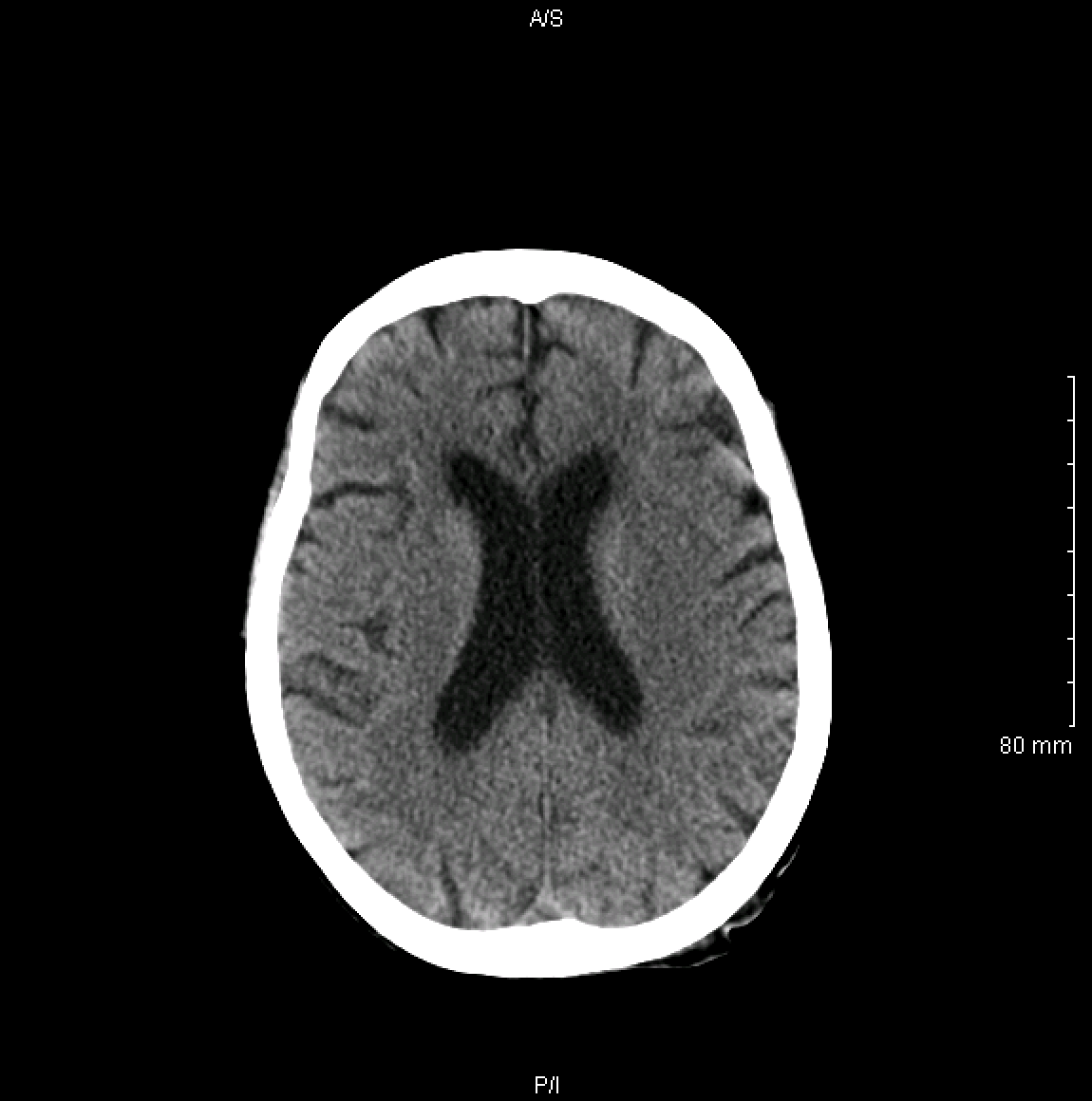
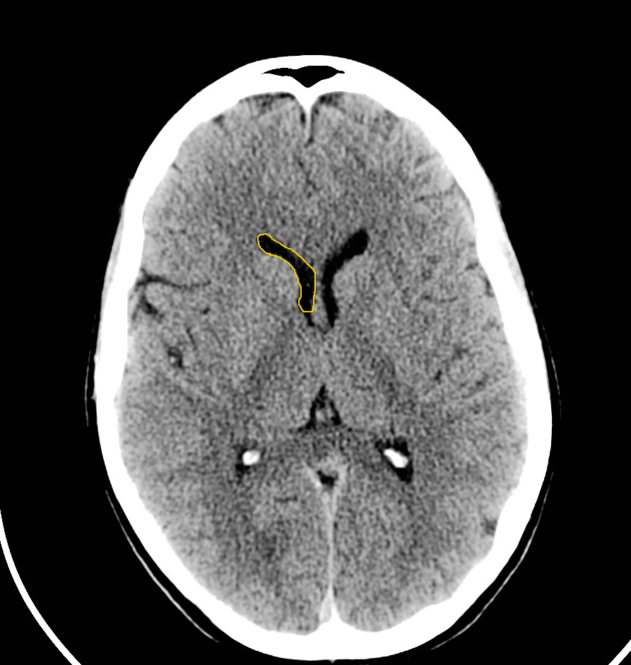
Non contrast axial ct head.

Ct scan anatomy of brain. Ct images of the brain are conventionally viewed from below as if looking up into the top of the head. Ct does not clearly show the anatomical borders of the lobes of the brain. Brain and face ct.
Ct scans are created using a series of x rays which are a form of radiation on the electromagnetic spectrum. Brain ct scans can provide more detailed information about brain tissue and brain structures than standard x rays of the head thus providing more data related to injuries andor diseases of the brain. This article lists a series of labeled imaging anatomy cases by system and modality.
This means that the right side of the brain is on the left side of the viewer. The anterior part of the head is at the top of the image. Angiogram coronal ct head.
Anatomy of the head on a cranial ct scan. Learn ct scan learn the diagnosis of ct and methods of computed tomography. Non contrast coronal ct head.
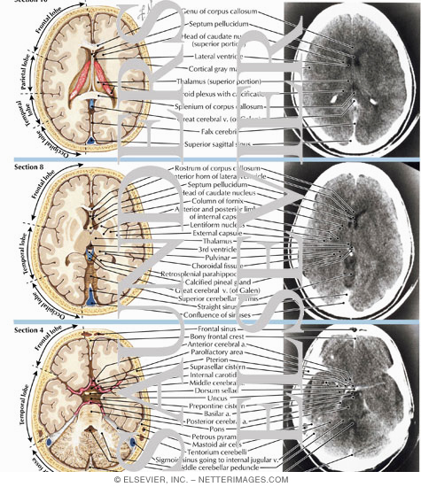
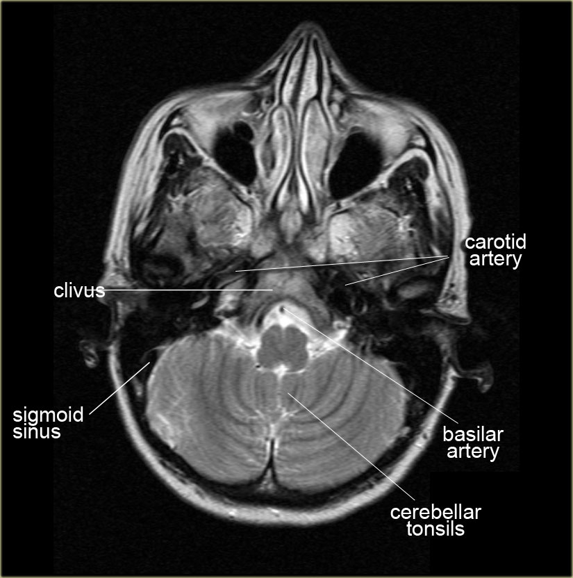
6 frontal bone 27 occipital bone 32 optic nerve 43 frontal sinus 45 sigmoid sinus 46 internal carotid artery. For this reason radiologists often refer to regions such as the parietal region or temporal region rather than lobes. Head ct scan intracranial ct scan a ct of the brain is a noninvasive diagnostic imaging procedure that uses special x rays measurements to produce horizontal or axial images often called slices of the brain.
The scanner emits x rays towards the patient from a variety of angles and the detectors in the scanner measure the difference between the x rays that are absorbed by the body and x rays that are transmitted through the body. Angiogram axial ct head. Anatomy of the head on a cranial ct scan.
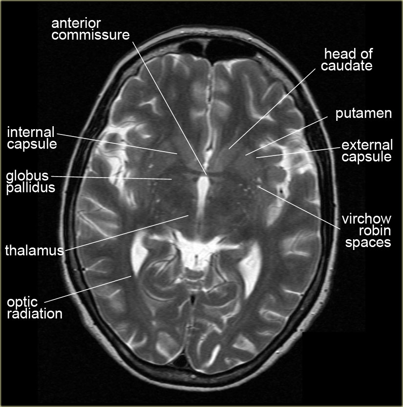
The lecture discussing the basic ct anatomy of the brain. Anatomy ct axial brain anatomy ct axial brain form no 1. Brain bones of cranium sinuses of the face.
Anatomy ct axial brain form no 19. What is a ct scan of the brain. Non contrast sagittal ct head.
 How To Read A Head Ct Emergency Medicine Newyork
 Normal Brain Anatomy As Demonstrated By Computerized
 Mri Basics
 Brain And Face Ct Interactive Anatomy Atlas
 Crash Ctscan Enlargments Brain Anatomy Radiology Neurology
 Ct Scan Cross Sectional Anatomy For Android Apk Download

 Ct Head Normal Anatomy
 Brain Imaging
 E Anatomy Radiologic Anatomy Atlas Of The Human Body
 The Radiology Assistant Brain Anatomy
 Figure 69 5 From How To Read A Head Ct Scan Semantic Scholar
 Test Yourself Ct Brain Quiz 2

 The Radiology Assistant Brain Anatomy
 Brain And Face Ct Interactive Anatomy Atlas
![]() Medical Imaging And Radiological Anatomy X Ray Ct Mri
Medical Imaging And Radiological Anatomy X Ray Ct Mri
 Brain And Face Ct Interactive Anatomy Atlas
 Head Ct Scan Procedure Radtechonduty
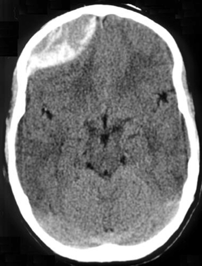
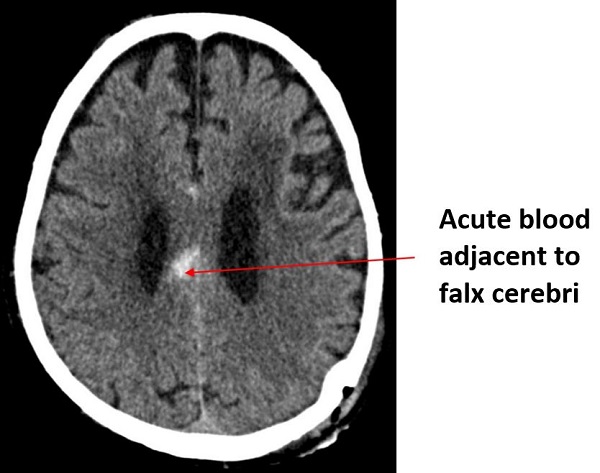
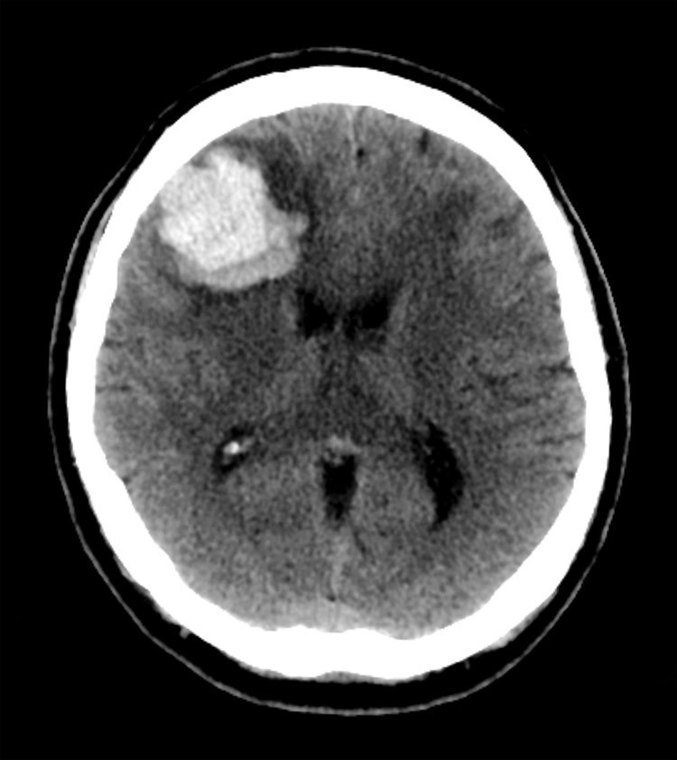
 Radiology Basics Head Pathology
 The Radiology Assistant Brain Anatomy
 Head Computed Tomography Scanning Background Indications
 The Radiology Assistant Brain Anatomy
![]() Medical Imaging And Radiological Anatomy X Ray Ct Mri
Medical Imaging And Radiological Anatomy X Ray Ct Mri
 Brain Imaging
 Brain And Face Ct Interactive Anatomy Atlas
 Brain Lobes Annotated Mri Radiology Case Radiopaedia Org
 Head Ct Interpretation Made Easy
Belum ada Komentar untuk "Ct Scan Anatomy Of Brain"
Posting Komentar