Coronary Sinus Anatomy
Knowledge of the anatomy of the coronary sinus. Chambers of the heart the body respectively and the coronary sinus draining blood from the heart itself.
Technical Aspects Of Coronary Sinus Catheterization Based On
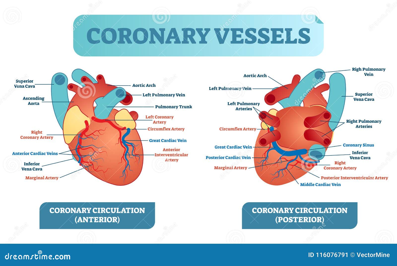
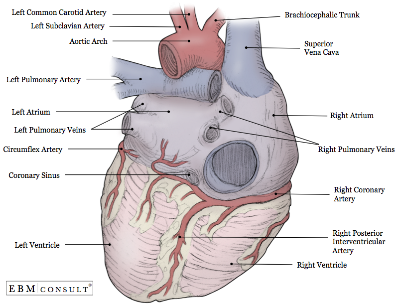
The coronary sinus is a collection of smaller veins that merge together to form the sinus or large vessel which is located along the hearts posterior rear surface between the left ventricle and left atrium.
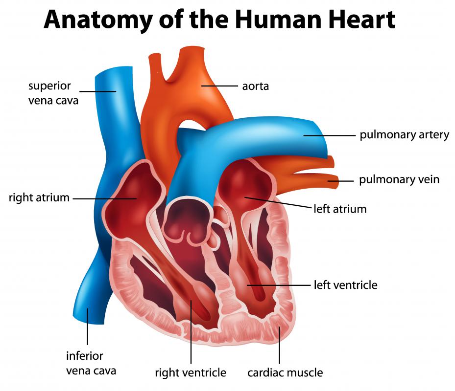
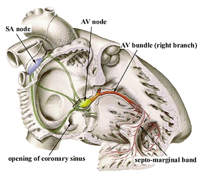
Coronary sinus anatomy. The coronary sinus empties directly into the right atrium near the conjunction of the posterior interventricular sulcus and the coronary sulcus crux cordis area located between the inferior vena cava and tricuspid valve. The tributaries of the greater and smaller cardiac veins or the thebesian veins. The coronary sinus courses along the posterior wall of the left atrium into the left atrioventricular groove.
85 of venous drainage occurs via the coronary sinus which is formed from the cardiac veins. Blood flows from the right atrium to the right ventricle. Coronary sinus anatomy the myocardium drains mainly by two groups of veins.
It returns blood from the heart muscle and is protected by a semicircular fold of the lining membrane of the auricle. Imaging of the coronary venous system has traditionally been. The other horn becomes part of the right atrium.
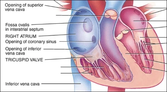
The coronary sinus drains into the right atrium at the coronary sinus orifice an opening between the inferior vena cava and the right atrioventricular orifice or tricuspid valve. This transition point is usually marked by the presence of intravenous valves which can obstruct catheter and lead placement. The right ventricle the right inferior portion of the heart is the chamber from which the pulmonary artery carries blood to the lungs.
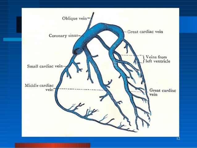
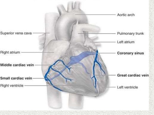
The great cardiac vein terminates in the coronary sinus a junction defined by the presence of the left atrial oblique vein. The circumference of the vein is larger than average and is big enough to allow blood to be deposited by most veins that enter the heart. The coronary sinus develops from one of the two sinus horns of the sinus venosum.
In human cardiovascular system. Normal anatomy and congenital abnormalities abstract. Coronary sinus gross anatomy.
Imaging of the coronary sinus. This atrial ostium can be partially covered by a thebesian valve although the anatomy of this valve is highly variable. The great cardiac vein runs with the lad the middle cardiac vein follows the pda.
Conventional coronary venous anatomy. The main vein of the greater venous system is the cs that runs in the posterior aspect of the coronary groove. Describe the normal anatomy of the cardiac venous system.
Another important branch is the middle cardiac vein.
 Great Cardiac Vein An Overview Sciencedirect Topics
 Coronary Sinus Ventricular Accessory Connections Producing
 Human Anatomy Mcqs Postgraduation Entrance Preparation 44
 Coronary Sinus Dr S Venkatesan Md
 Coronary Sinus Posterior View The Anatomy Of The Heart
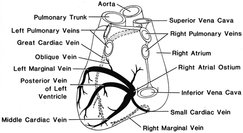 Anatomy Atlases Illustrated Encyclopedia Of Human Anatomic
Print Anatomy 2 Lab Flashcards Easy Notecards
 Figure 1 From The Anatomy Of The Coronary Sinus Venous
:background_color(FFFFFF):format(jpeg)/images/library/8798/coronary-arteries-and-cardiac-veins_english.jpg) Coronary Arteries And Cardiac Veins Anatomy And Branches
Coronary Arteries And Cardiac Veins Anatomy And Branches
 Anatomical Relationship Between The Coronary Sinus Cs And
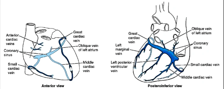 Thorax Venous Structure Coronary Veins Ranzcrpart1 Wiki
Coronary System Tutorial What Is The Coronary System
 E Coronary Sinus After Passing Through The Capillary Beds
Coronary Anomalies What The Radiologist Should Know
 Coronary Sinus Stock Photos Coronary Sinus Stock Images
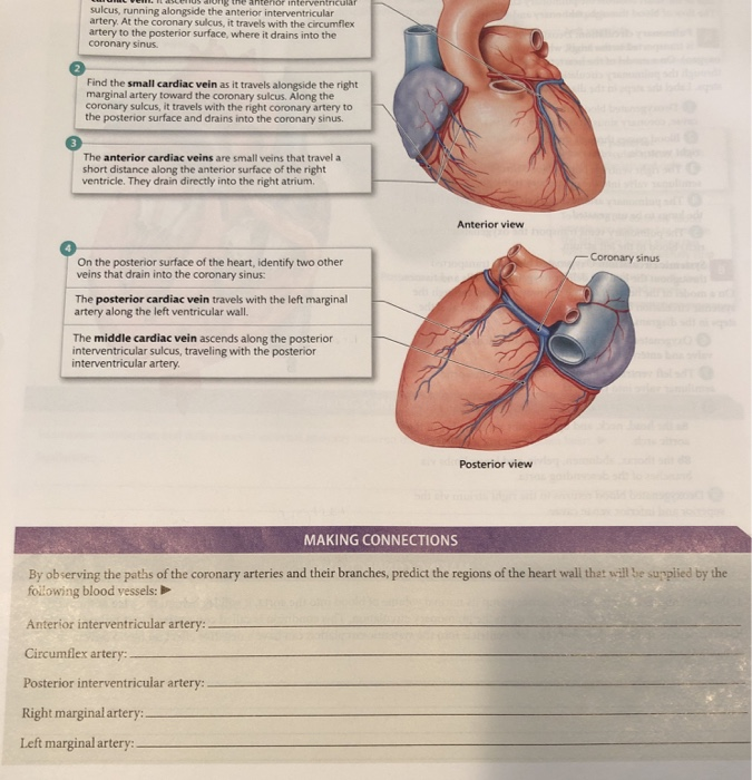 Solved Sulcus Running Alongside The Anterior Interventri
 Great Cardiac Vein An Overview Sciencedirect Topics
 Types Of Cs Coronary Sinus Anatomy According To M Von
 Cardiac Venous System Venous And Lymphatic Diseases
 What Is The Coronary Sinus With Pictures
 Right Coronary Artery An Overview Sciencedirect Topics
 Coronary Sinus Stock Illustrations 22 Coronary Sinus Stock
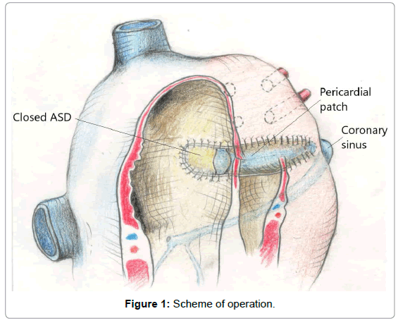 A Complete Anatomical Correction Of An Atrial Septal Defect
 Coronary Sinus Dr S Venkatesan Md



Belum ada Komentar untuk "Coronary Sinus Anatomy"
Posting Komentar