Anatomy Of Thoracic Cage
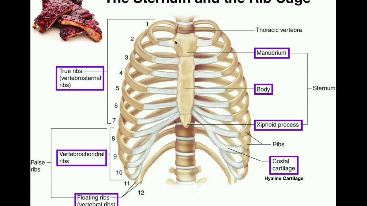
This 3d anatomy tutorial covers the basic bony framework of the thoracic wall. The thoracic cage is formed by the 12 pairs of ribs with their costal cartilages and the sternum.
 Rib Cage Wikipedia
There are five muscles that make up the thoracic cage.
Anatomy of thoracic cage. It encloses the thoracic cavity which contains the lungs. There are some other muscles that do not comprise the thoracic wall but do attach to it. It is formed by the 12 thoracic vertebrae 12 pairs of ribs and associated costal cartilages and the sternum.
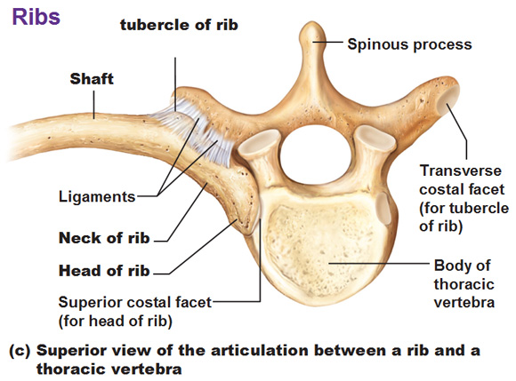
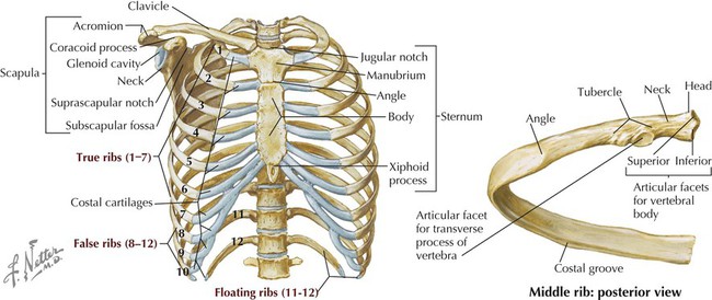
The ribs are anchored posteriorly to the 12 thoracic vertebrae t1t12. These include the pectoralis. These muscles act to change the volume of the thoracic cavity during respiration.
An inhalation is accomplished when the muscular diaphragm at the floor of the thoracic cavity contracts and flattens while the contraction of intercostal muscles lift the rib cage up and out. The human rib cage is a component of the human respiratory system. Osteology of the thoracic cage.
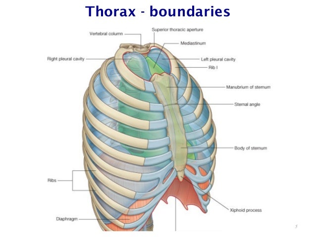
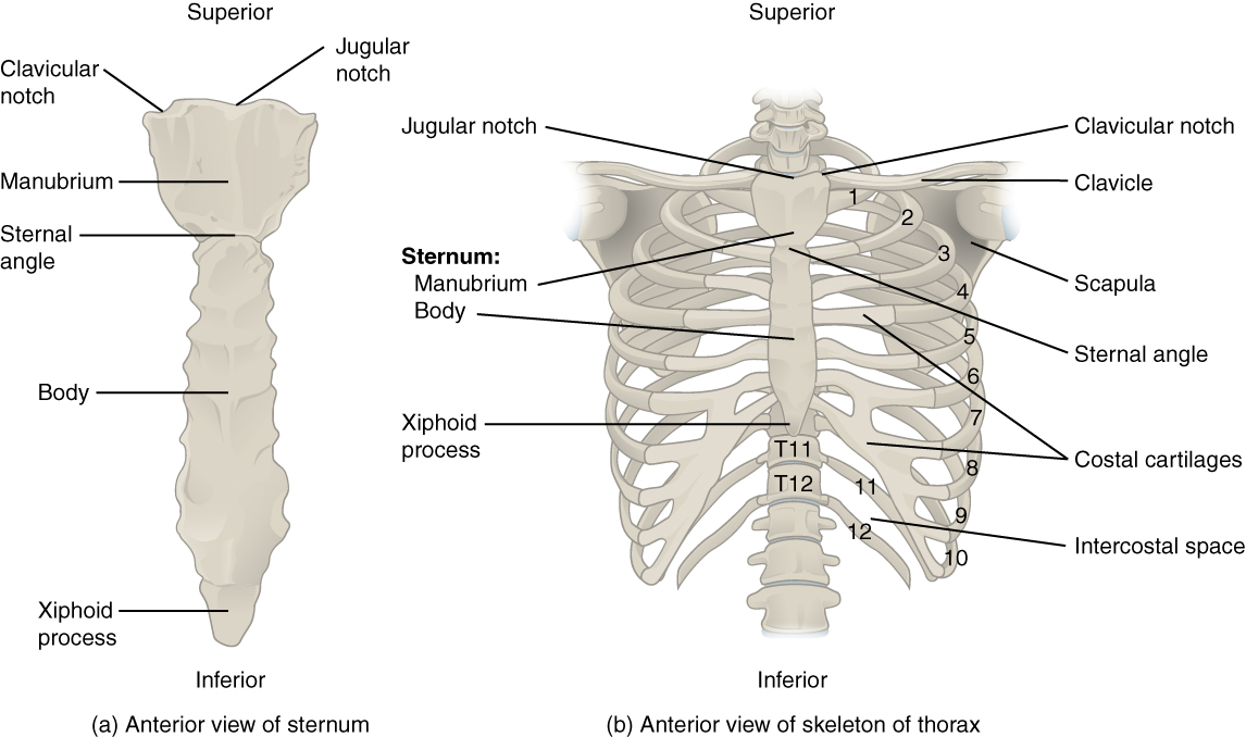
The thoracic cage rib cage is the skeleton of the thoracic wall. It consists of the 12 pairs of ribs with their costal cartilages and the sternum figure 732. Ribs and costal cartilages duration.
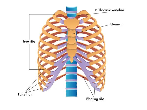
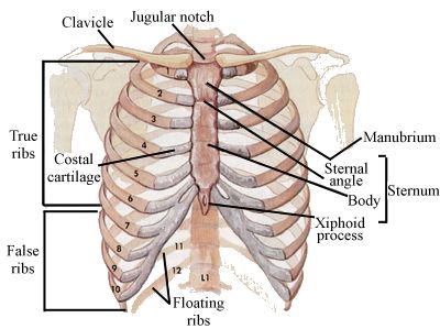
The thoracic cage functions to protect the heart and lungs. The ribs are attached posteriorly to the 12 thoracic vertebrae and most are anchored anteriorly either directly or indirectly to the sternum. The thoracic cage functions to protect the heart and lungs.
This video focusses on the naming of bones of the rib cage and the bony structures associated with it including the sternum. The thoracic cage is formed by the 12 pairs of ribs with their costal cartilages and the sternum. The intercostals external internal and innermost subcostals and transversus thoracis.
A tutorial will be following soon on the musculature of the thoracic wall to complement this. Video number anatomy 1 33. The thoracic cage rib cage forms the thorax chest portion of the body.
Anatomy thorax overview ribs sternal angle pleura and pneumothorax armando hasudungan. The thoracic cage takes the form of a domed bird cage with the horizontal bars formed by ribs and costal cartilages. The ribs are attached posteriorly to the 12 thoracic vertebrae and most are anchored anteriorly either directly or indirectly to the sternum.
The thoracic cage protects the heart and lungs.
 Anatomy The Sternum Rib Cage Vertebrae
 Anterior View Of The Anatomy Thoracic Cage Photo Axial
 Rib Cage Anatomy Britannica
![]() Human Skeleton Structure Skull Spine Rib Cage Pelvis Joints
Human Skeleton Structure Skull Spine Rib Cage Pelvis Joints
 Thoracic Cage
 Chapter 2 Anterior Thoracic Wall The Big Picture Gross
 Unit Iv
 Basic Anatomy Thoracic Cage Powerpoint Recording
 ᐈ Rib Cage Silhouette Stock Vectors Royalty Free Rib Cage

 Basic Biomechanics Of The Thoracic Spine And Rib Cage
 Rib Cage Wikipedia
 Thorax Basicmedical Key
 Skeletal Series Part 5 The Human Rib Cage These Bones Of Mine
 Rib Cage Human Skeleton Human Body Anatomy Thoracic Cage
 7 4 The Thoracic Cage Anatomy And Physiology

 Human Rib Cage Anatomy White Background Plain Background
 Thoracic Wall And Breast Illustrations
 Pectus Carinatum Pc Is An Anterior Chest Wall Deformity

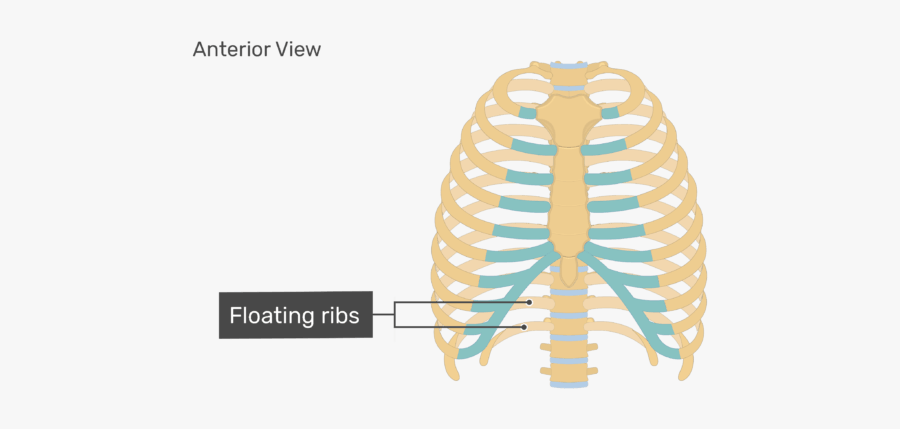
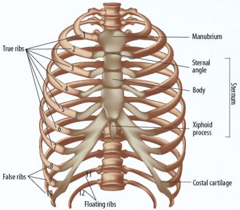
Thoracic Cage Ribs Fontanelles
 Unit Iv
 Skeleton Clinical Anatomy With Freddo At Massachusetts
 Eps Vector Rib Cage Bones Human Skeletal System Anatomy
 The Thoracic Cage The Rib Cage
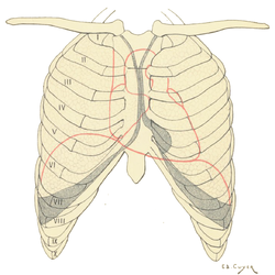
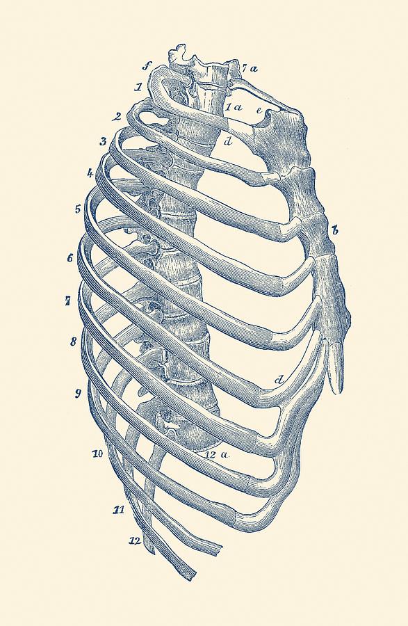 Rib Cage Diagram Vintage Anatomy Print
 1 Schematic Illustration Of The Anatomy Of The Thoracic Cage
 Dbcs Anatomy Thoracic Cage Diagram Quizlet
Belum ada Komentar untuk "Anatomy Of Thoracic Cage"
Posting Komentar