Anatomy Of The Large Intestine
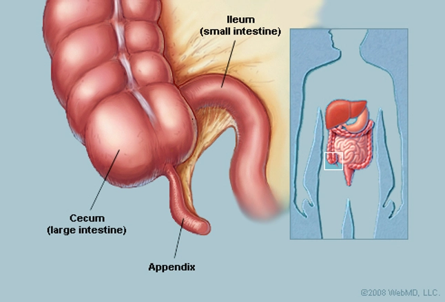
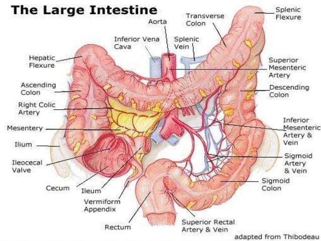
Large intestine anatomy and physiology as mentioned earlier the large bowel starts from the point where the small intestine ends. The ileocecal valve located at the opening between the ileum and the large intestine controls the flow of chyme from the small intestine to the large intestine.
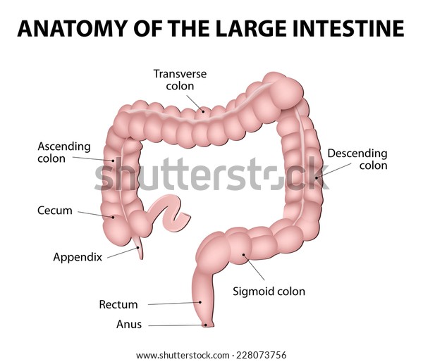 Anatomy Large Intestine Medical Illustration Human Stock
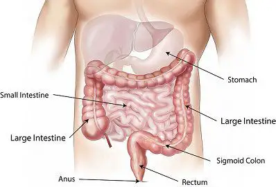
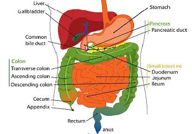
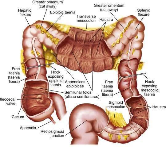
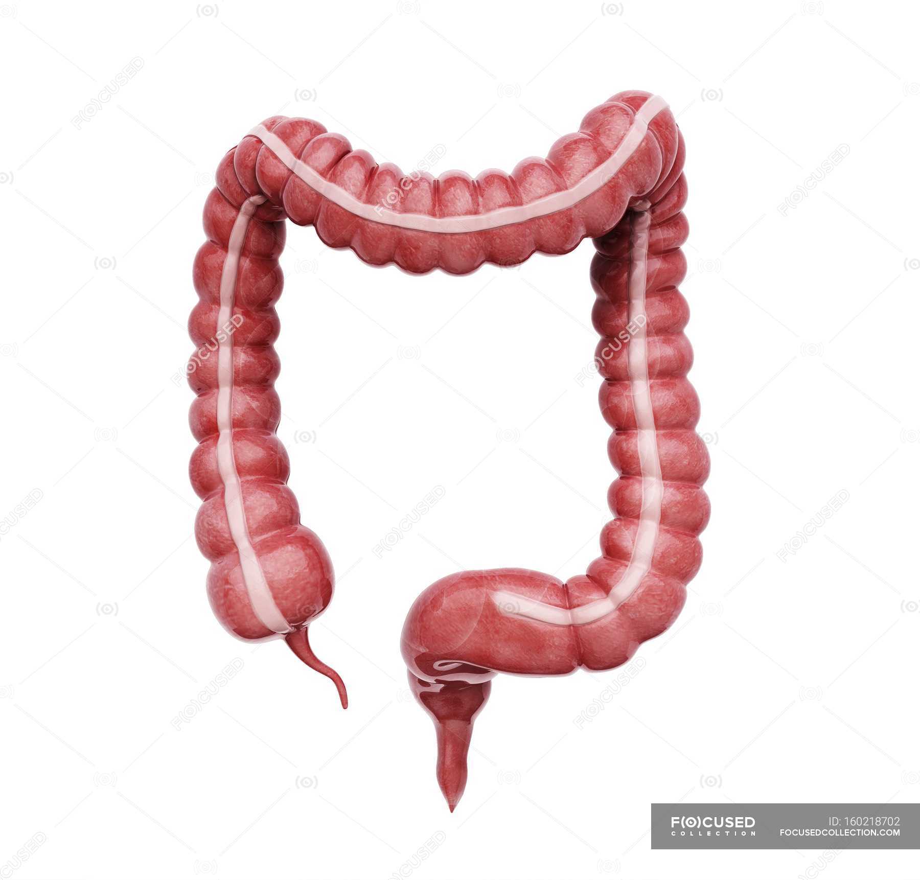
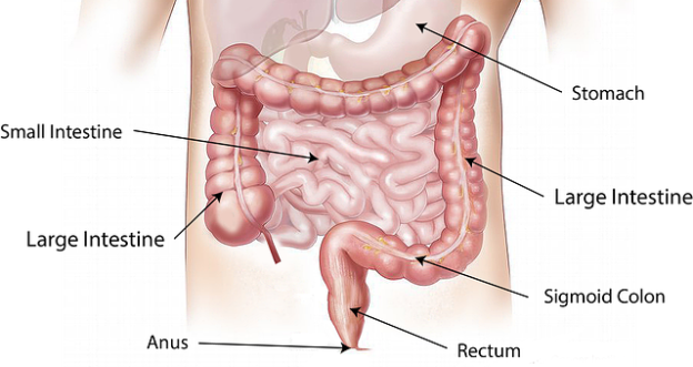
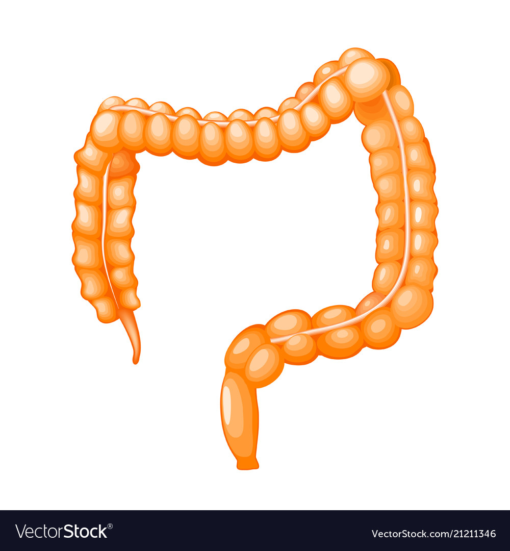
The large intestine consists of the cecum ascending colon transverse colon descending colon and sigmoid colon.
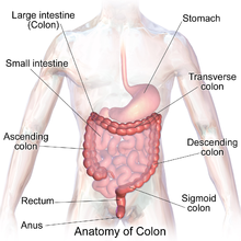
Anatomy of the large intestine. The large intestine consists of the cecum colon rectum and anal canal. The large intestine consists of the cecum appendix ascending colon transverse colon descending colon rectum and anal canal. The invaginations are called the intestinal glands or colonic crypts.
The large intestine is responsible for processing indigestible food material chyme after most nutrients are absorbed in the small intestine. The cecum the colon the rectum and the anus. The large intestine is about 5 feet 15 m in length and 25 inches 6 7 cm in diameter in the living body but becomes much larger postmortem as the smooth muscle tissue of the intestinal wall relaxes.
Gross and microscopic anatomy of the large intestine the large intestine is that part of the digestive tube between the terminal ileum and anus. The wall of the large intestine is lined with simple columnar epithelium with invaginations. The large intestine or large bowel is the last part of the digestive system in vertebrate animals.
Depending on the species ingesta from the small intestine enters the large intestine through either the ileocecal or ileocolic valve. Although shorter than the small intestine in length the large intestine is considerably thicker in diameter thus giving it its name. Its function is to absorb water from the remaining indigestible food matter and then to pass the useless waste material from the body.
The large intestine is named for its relatively large diameter not its length. To be more precise it starts from the right iliac region of the pelvis. The colon crypts are shaped like microscopic thick walled test tubes with a central hole down the length of the tube the crypt lumen.
The large intestine is subdivided into four main regions.
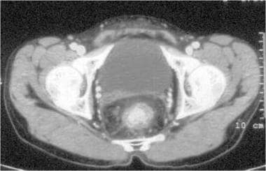 Large Intestine Anatomy Gross Anatomy Histology Natural
 Vector Illustration Large Intestine Anatomy Eps Clipart
 Digestive System 4 Large Intestine Anatomy Physiology
 Large Intestine Human Anatomy Canvas Print
 Human Intestines Interactive Anatomy Guide
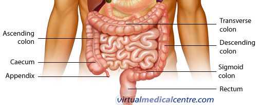 Gastrointestinal System Anatomy Healthengine Blog
 Large Intestine Images Stock Photos Vectors Shutterstock
 Part Of Large Intestine Isolated On A White Background Medical
 Appendix Anatomy Appendix Picture Location Definition
Item Detail Anatomy Pad Colon Large Intestine And Rectum
 Difference Between Small Intestine And Large Intestine With
 The Large Intestine Human Anatomy
 Large Intestine Structure And Function Preview Human Anatomy Kenhub
 Difference Between Small Intestine And Large Intestine With
 Anatomy Of The Large Intestine Doctor Stock
 Colonoscopy Anatomy Of The Colon Healthlink Bc
 Gross Anatomy Of The Large Intestine Purposegames
 Human Digestive System Secretions Britannica
 Anatomy Histology Embryology And Developmental Anomalies
Large Intestine Anatomy And Physiology
 The Colon What It Is What It Does Ascrs
 Anatomy Of Normal Large Intestine White Background Plain
 Large Intestine Large Bowel Anatomy Functions And Pathology
 Anatomy And Physiology Of The Large Intestine


Belum ada Komentar untuk "Anatomy Of The Large Intestine"
Posting Komentar