Hamstring Tendon Anatomy
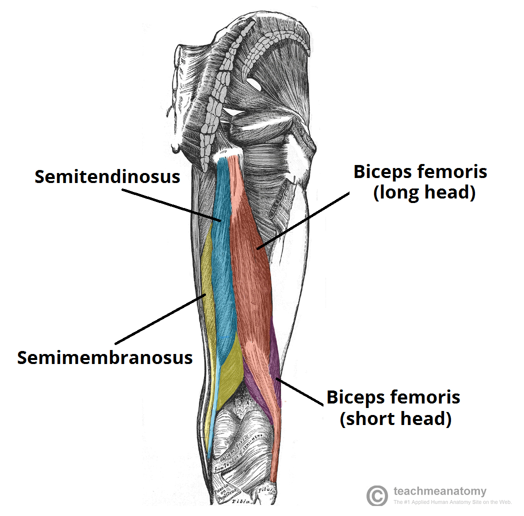
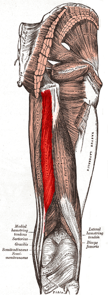
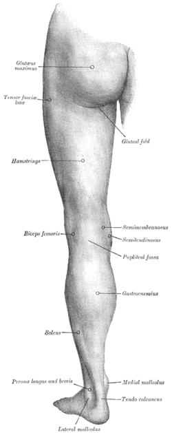
The most medial muscle the semimembranosus. The hamstrings are comprised of the semimembranosus semitendinosus and the biceps femoris muscles.
 Muscles Of The Posterior Thigh Hamstrings Damage
Sudden sharp pain may be felt at the time of injury with possible swelling and soreness.
Hamstring tendon anatomy. As group these muscles act to extend at the hip and flex at the knee. Highly folded membranes at the muscle tendon interface increase junctional surface area an adaptation designed to dissipate energy 27. Muscle will participate in flexion of the knee.
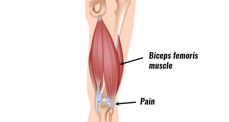
The muscle belly of the semitendinosus is the most cepahalad muscle of the hamstring complex. They consist of the biceps femoris semitendinosus and semimembranosus which form prominent tendons medially and laterally at the back of the knee. A full or partial rupture of the hamstring muscle tendons can occur at the point where they insert into the back of the knee.
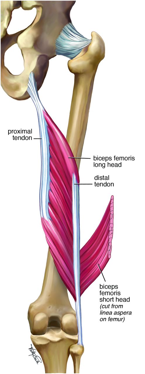
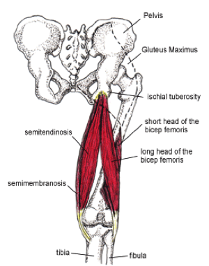
Muscles should originate from ischial tuberosity. The lateral hamstring is the biceps femoris made up of 2 parts a short head and long head and the medial hamstrings are the semitendinosus joins the sartorius muscle and gracilis muscle at the pes anserinus on the tibia and the semimembranosus the largest hamstring muscle. The hamstring muscles have their origin where their tendons attach to bone at the ischial tuberosity of the hip often called the sitting bones and the linea aspera of the femur.
The hamstring tendons flank the space behind the knee. The common criteria of any hamstring muscles are. Hamstring tendon anatomy the hamstring muscle group is comprised of three separate muscles.
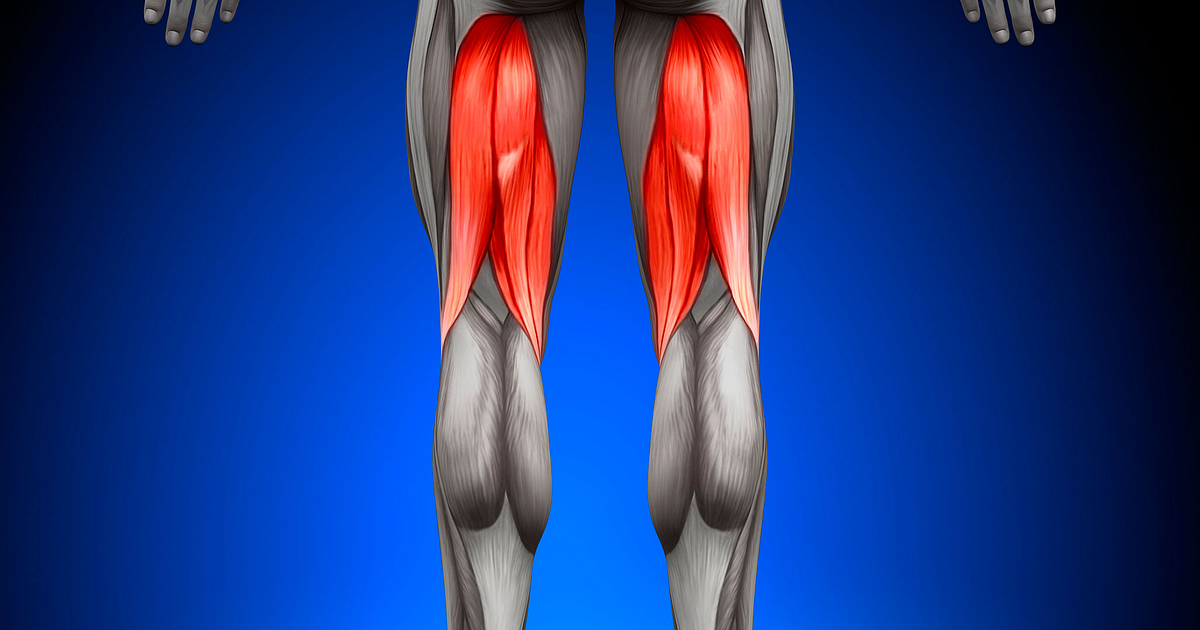
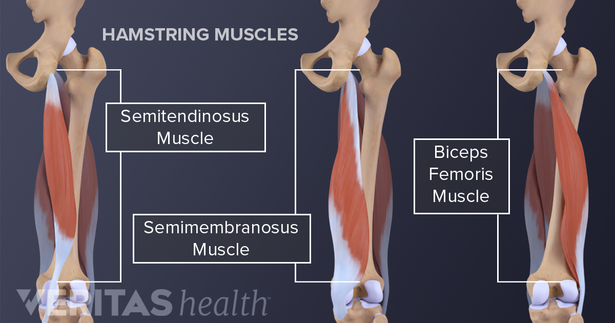
The biceps femoris short and long head semimembranosus and the semitendinosus see figure 1. Anatomy of the hamstring muscles. The muscles in the posterior compartment of the thigh are collectively known as the hamstrings.
Muscles will be innervated by the tibial branch of the sciatic nerve. Hamstring muscle injury typically occurs in the region of the mtj which as opposed to being a distinct point is really a 1012 cm transition zone in which myofibrils contribute to form the tendon. The hamstrings are the muscles of the posterior compartment of the thigh and include the.
At the top of the muscle group while the short head of biceps femoris attaches to the femur all the other hamstring muscles share a common point of origin on the ischial tuberosity sitting bones of the pelvis. Anatomy of the hamstring muscles there are actually three hamstring muscles at the back of each of your thighs. Semitendinosus semimembranosus and biceps femoris with its long and short heads.
Muscles should be inserted over the knee joint in the tibia or in the fibula. The biceps femoris and semitendinosus arise from a common tendon along the posteromedial aspect of the ischial tuberosity. Andrew murphy and dr craig hacking et al.
Hamstrings The Muscle At The Back Of The Upper Thigh
 Strains Of The Hamstring Strains Occur In Muscles And
How Serious Is A Hamstring Injury Orthopedic Spine Therapy
 Acl Hamstring Tendon Graft Reconstruction Knee Injuries
 Serious Thigh Muscle Strains Beware The Intramuscular
Patient Education Concord Orthopaedics
 Hamstring Tendonitis Or Hamstring Syndrome Zion Physical
 Hamstring Graft For Anterior Cruciate Ligament Repair
 Hamstring Injury Causes Symptoms Recovery Time Treatment
 A Pain In The Rear High Hamstring Tendinitis Runner S World
 Pulled Hamstring Causes Symptoms Recovery And Treatment
 Hamstring Injuries The Hughston Clinic
Search Rupture Of Hamstring Tendon With Edema
 Hamstring Tendonitis Symptoms Causes Treatment Exercises
 Proximal Hamstring Tendinopathy Physiopedia
Vivian Grisogono About The Calf
 Hamstring Strain And Pulled Hamstring Injury Treatment
 You Have Nine Hamstring Muscles
 Hamstring Tendonitis Hamstring Injuries Physioadvisor
 Chronic High Proximal Hamstring Tendinopathy
 Avoiding The Sniper Shot Hamstring Pulls Sparta Science
 Sitbone Pain From Yoga Asana Love Yoga Anatomy


Belum ada Komentar untuk "Hamstring Tendon Anatomy"
Posting Komentar