Anatomy Rotator Cuff
It is innervated by the upper and lower subscapular nerves. This holds your humerus in place and keeps your upper arm stable.
 Rotator Cuff Pain Tears And Other Injures Treatments
Tests for a rotator cuff tear may include.
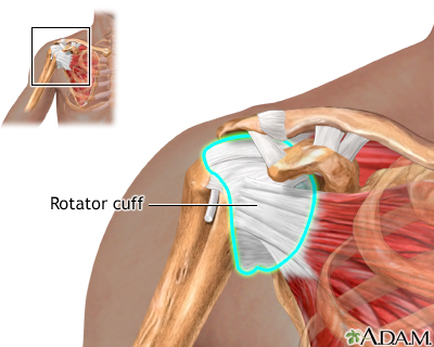
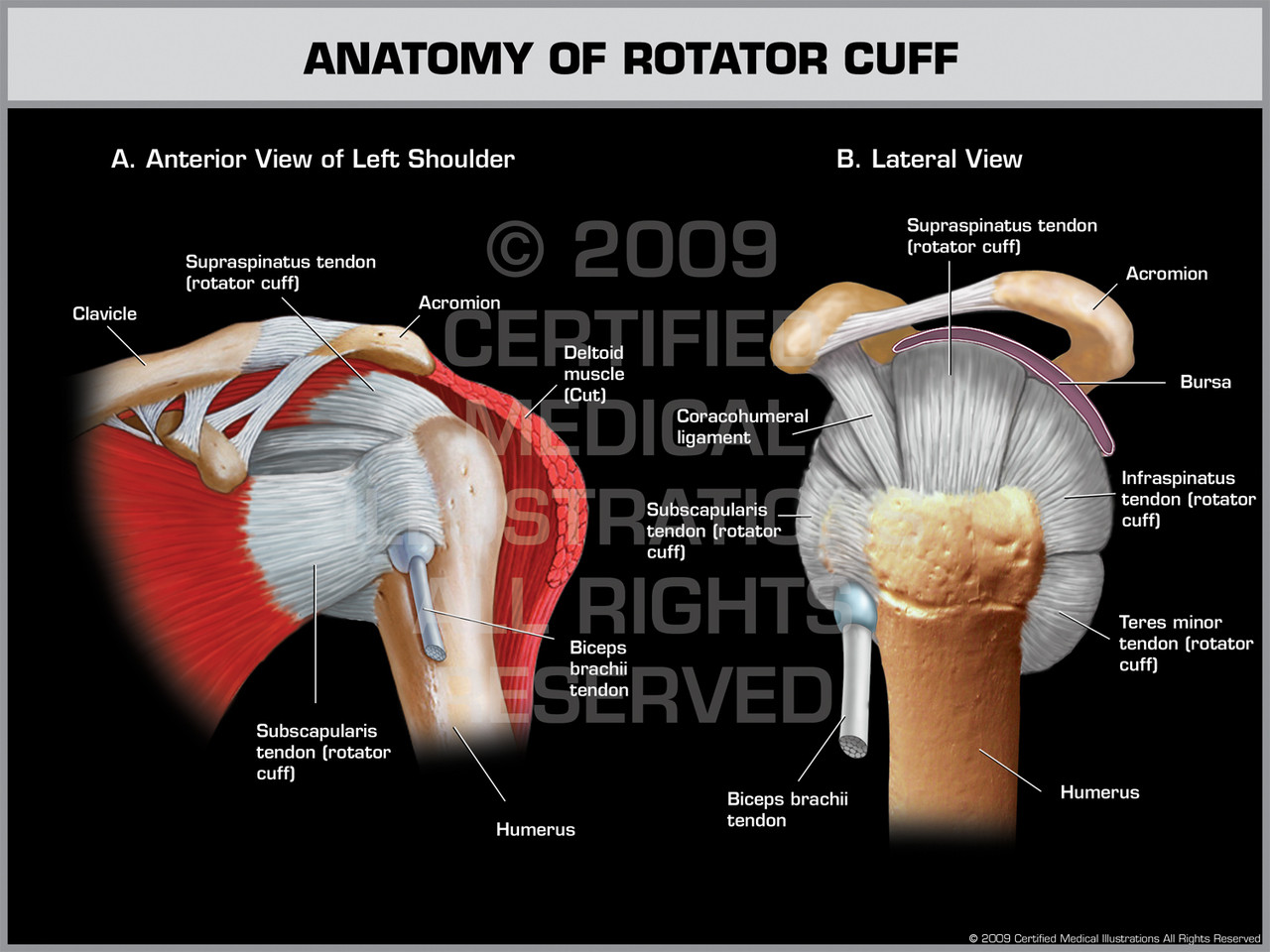
Anatomy rotator cuff. The joint where the upper bone humerus of the arm meets the shoulder scapula and acromion process is called the rotator cuff. This is the main muscle that lets you rotate and extend your shoulder. Its function is related to the glenohumeral joint where the muscles of the cuff function both as the executors of the movements of the joint and the stabilization of the joint as well.
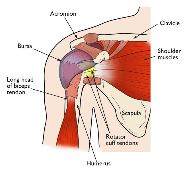
Together with the joint capsule ligaments and labrum the rotator cuff muscles are important dynamic stabilizers and movers of the shoulder joint. Can be localized to anterior lateral aspect of the shoulder with referred pain down the upper arm lateral aspect. The most common signs of rotator cuff injuries are.
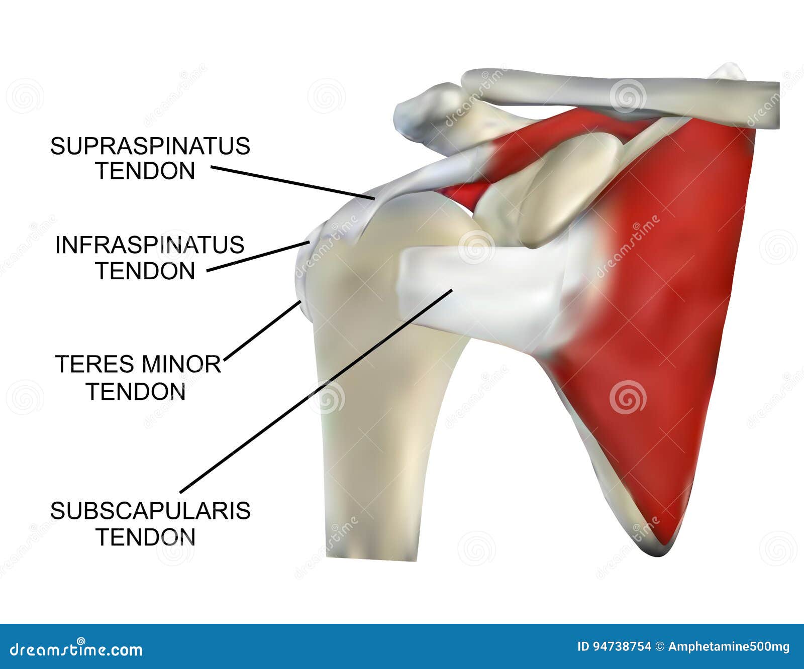
The rotator cuff are a group of muscles which are important in supporting the glenohumeral joint. The four muscles are the supraspinatus muscle the infraspinatus muscle teres minor muscle and the subscapularis muscle. The rotator cuff is the most vulnerable part of the shoulder and is where most shoulder injuries occur.
In anatomy the rotator cuff is a group of muscles and their tendons that act to stabilize the shoulder and allow for its extensive range of motion. They are important in shoulder movements and maintaining stability of this joint. Skip repeated content.
Arthrogram a special type of x ray that uses dye injected into a joint to more clearly see detail in the tendons and muscles. In the human body the rotator cuff is a functional anatomical unit located in the upper extremity. Painful range of motion.
Anatomy of rotator cuff the subscapularis arises from the anterior aspect of the scapula and attaches over much of the lesser tuberosity. Of the seven scapulohumeral muscles four make up the rotator cuff. This is the smallest rotator cuff.
Mri magnetic resonance imaging. Each one of these muscles is part of the rotator cuff and plays an important role. The rotator cuff is made up of four muscles whose tendons come together to form a covering around the head of the humerus upper arm bone and top of the shoulder.
They are important in shoulder movements and maintaining stability of this joint. Pain may or may not be present.
 Axis Scientific Anatomy Model Of Muscled Shoulder Joint Shows Complete Shoulder Musculature From Rotator Cuff To Subscapular Muscles Comes On
Anatomy Of A Rotator Cuff Tear Archives Arizona Institute
 Shoulder Injury Case Settlement Values For Torn Rotator
 Anatomy Of The Rotator Cuff Muscles Stock Illustration
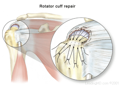
 Rotator Cuff Repair Series Normal Anatomy Medlineplus
 Shoulder Pain Learn About The Rotator Cuff Salvation Wellness
 Rotator Cuff Injuries Treatment How To Manage The Pain Heal
 Rotator Cuff Injury Shoulder Specialist Chicago
Shoulder Impingement Syndrome Cleveland Clinic
 Rotator Cuff Tears Orthoinfo Aaos
 Rotator Cuff Images Stock Photos Vectors Shutterstock
 Rotator Cuff Muscles Of The Left Shoulder Stock Trial Exhibits
 Shoulder Replacement Recovery Rotator Cuff Anatomy Image
 Exam Series Guide To The Shoulder Exam Canadiem
10 Best Exercises To Strengthen Your Rotator Cuff Builtlean
 Mayo Clinic Q And A Treating Rotator Cuff Tears Mayo
 Cables Crescents And Suspension Bridges The Unique Anatomy
 What Is The Rotator Cuff And Why It S Important To Shoulder
 Rotator Cuff Tears Surgical Treatment Options Jewett
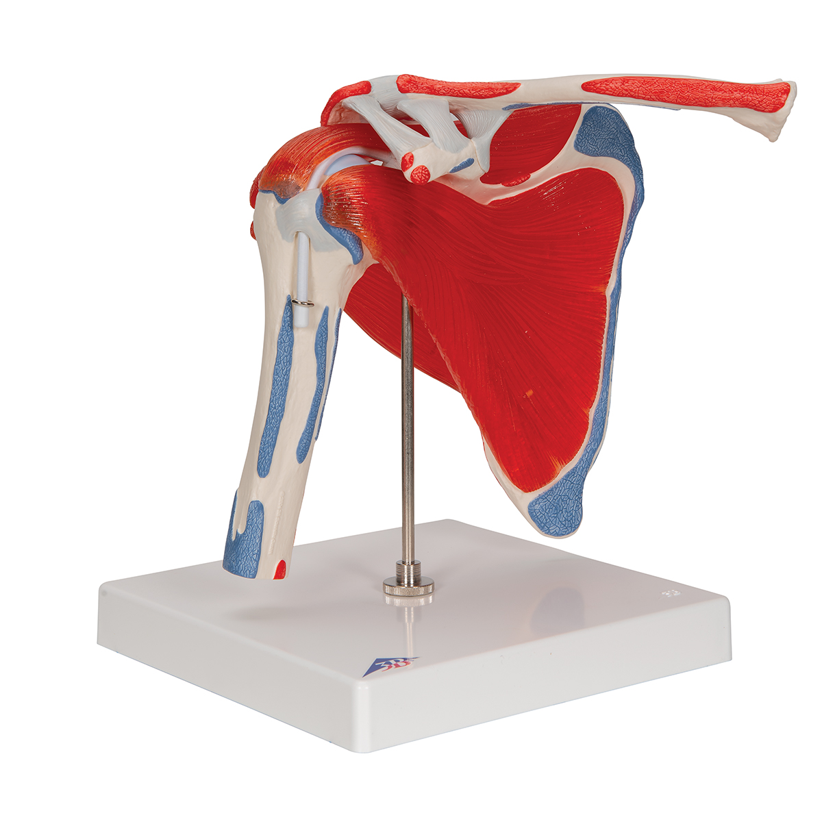 Anatomical Teaching Models Plastic Human Joint Models
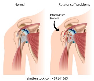 Rotator Cuff Images Stock Photos Vectors Shutterstock
 Shoulder Joint Anatomy Model With Rotator Cuff
 Pin By Heather Wagner On Our Bodies Shoulder Anatomy
 Rotator Cuff Anatomy Overview Physiostrength
 What Are The Causes Of Rotator Cuff Pathology
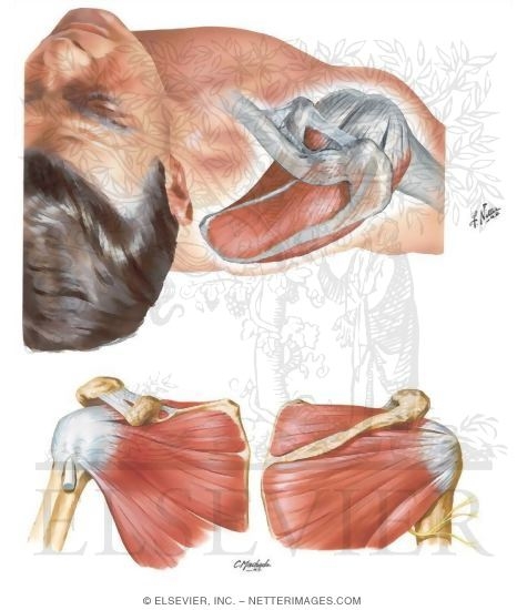
Belum ada Komentar untuk "Anatomy Rotator Cuff"
Posting Komentar