Thigh Anatomy Mri
Related posts of thigh muscle anatomy mri eye diagram muscles anatomy. Stanford msk mri atlas radlex.
 Fascial Compartments Of Thigh Wikipedia
Check for errors and try again.

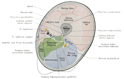
Thigh anatomy mri. Stanford bone tumor bayesian network issssr msk lectures for residents ocad msk cases from around the world stanford msk mri atlas has served almost 800000 pages to users in over 100 countries. Eye diagram muscles anatomy 16 photos of the eye diagram muscles anatomy diagram back muscles diagram leg muscles diagram of eye anatomy diagram of muscles in arm diagram of muscles in lower back diagram of muscles in the knee diagram of muscles in the neck human muscles diagram back muscles diagram leg muscles. Muscles of the thigh muscle origin insertion nerve supply sartorius anterior superior iliac spine and adjacent area below medial surface of the tibia.
Posterosuperior surface of the rim of the acetabulum through the patellar ligament to. 2 vastus medialis intermedius muscles. Anterior inferior iliac spine.
1 vastus lateralis muscle. Near the tuberosity and neighboring fascia femoral rectus femoris straight head. Injuries such as anterior cruciate ligament meniscus and rotator cuff tears are all easily diagnosed when there is a firm understanding and knowledge of human anatomy.
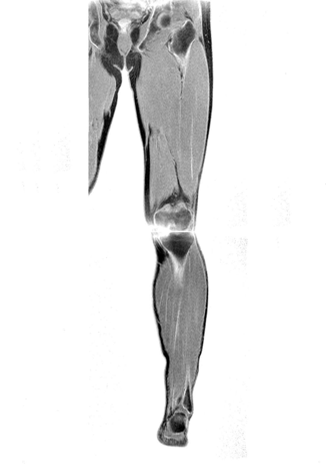
With an axial spin echo t1 weighted acquisition covering the entire human leg. Anatomy of the thigh. A magnetic resonance imaging mri was performed on a healthy subject.
Positioning for mri upper legs position the patient in supine position with feet pointing towards the magnet feet first supine position the patient over the spine coil and place the body coils over the thighs anterior superior iliac spine down to knee joints. Unable to process the form. Thigh refers to the portion of the lower limb between the hip and knee joints.
Note that in an anatomical context leg refers to the portion between the knee and ankle joints and not to the entire lower limb. Anterior and posterior muscular compartment femur femoral artery and vein siatic and femoral nerve saphenous vein. About anatomy mri magnetic resonance imaging is particularly well suited for the medical evaluation of the musculoskeletal msk system including the knee shoulder ankle wrist and elbow.
 Untitled Document


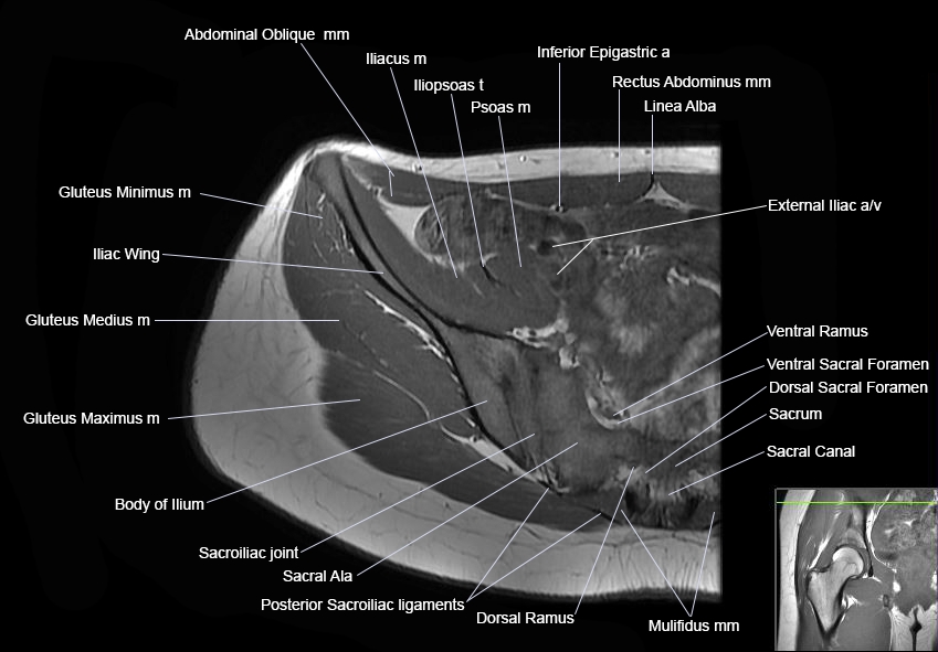

 Module 2 Lower Extremity Orthopedic Imaging
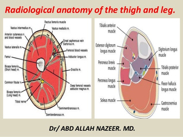 Presentation1 Pptx Radiological Anatomy Of The Thigh And Leg
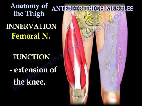 Anatomy Of The Thigh Everything You Need To Know Dr Nabil Ebraheim
 Module 2 Lower Extremity Orthopedic Imaging

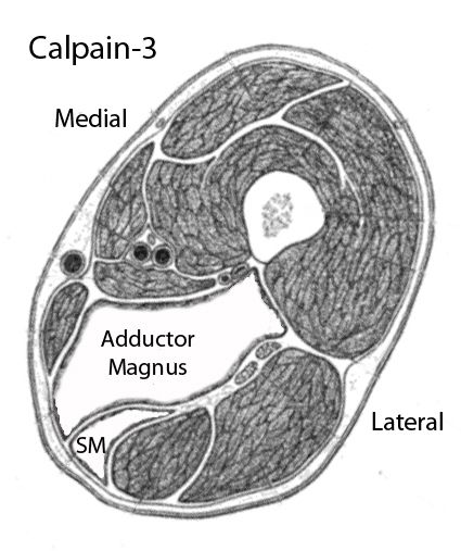 Muscle Mri

 X Rays Ct Scans And Mris Orthoinfo Aaos
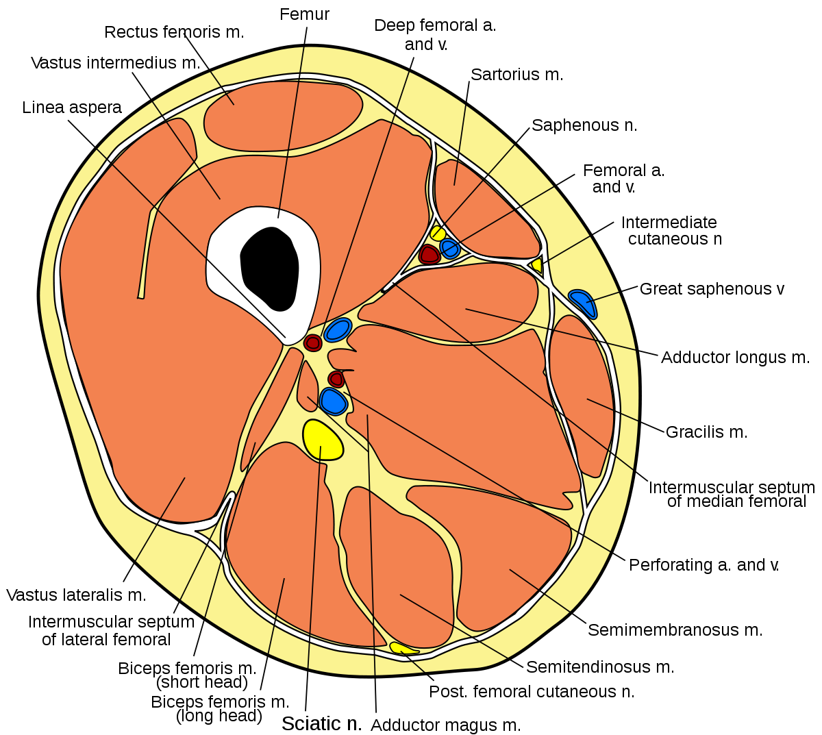 Lateral Intermuscular Septum Of Thigh Wikipedia
 K Anatomy
 Figure 4 From Normal Mr Imaging Anatomy Of The Thigh And Leg

Vastus Lateralis Anatomy Orthobullets
![]() Diagram Pictures Neurovasculature Of The Hip And The
Diagram Pictures Neurovasculature Of The Hip And The
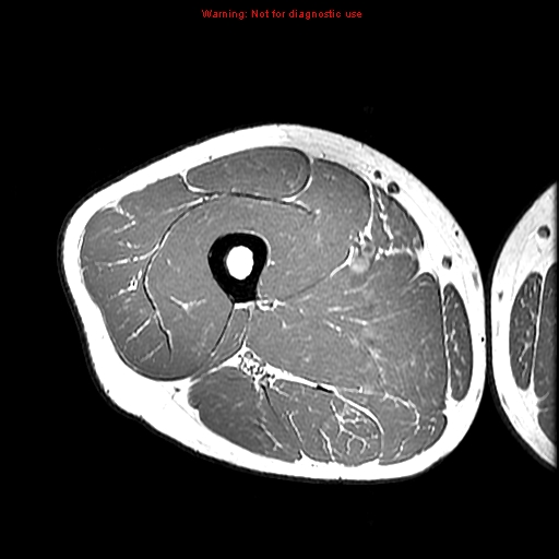
![]() T1 Axial Transverse Mri Of Thigh A Distal Insert Image
T1 Axial Transverse Mri Of Thigh A Distal Insert Image
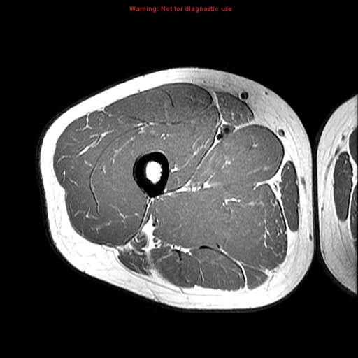
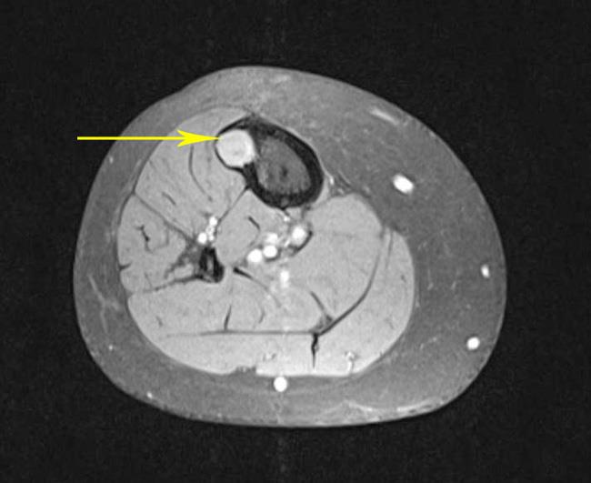
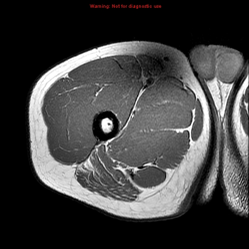
 Thigh Muscles Cross Sectional Anatomy Radiology Case
 Lower Extremity Mri Of Anatomical Atlas
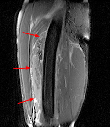
 Quadriceps Muscle Contusion Physiopedia

 Anatomy Thigh Axial Stress Fractures 78 Steps Health Journal
 Presentation1 Pptx Radiological Anatomy Of The Thigh And Leg
 Mri Thigh Calf Anatomy Normal Anatomy Dr Ahmed Eisawy

Belum ada Komentar untuk "Thigh Anatomy Mri"
Posting Komentar