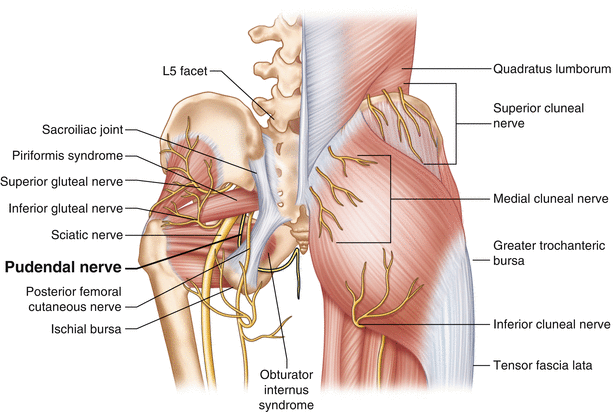
Pudendal Nerve Anatomy
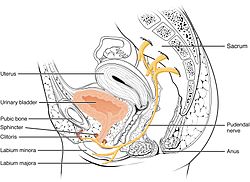
Anatomy of the pudendal nerve. It carries sensory information sensation from the external genitalia and the skin around the anus and perineum.
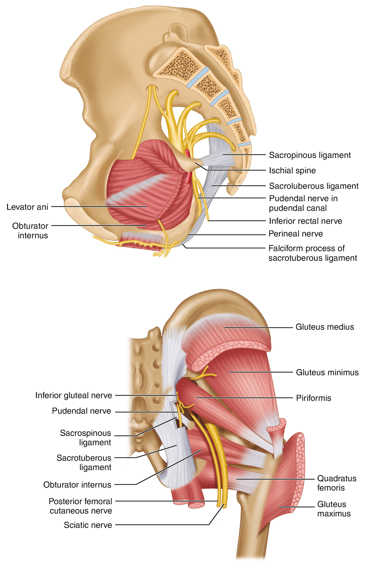
 Pudendal Nerve Anatomy Gray S Anatomy 2011 Download
The pudendal nerve emerges from the s2 s3 and s4 roots ventral rami of the sacral plexus.

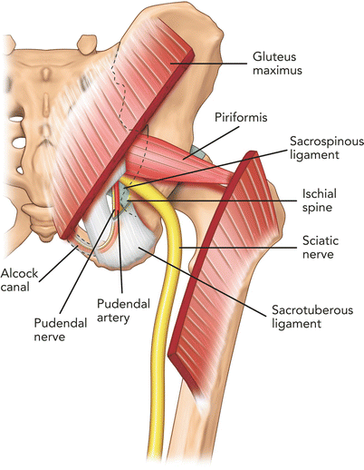
Pudendal nerve anatomy. The nerve extends from the sacral plexus through the pudendal canal the perineum and the gluteal area. The pudendal nerve is formed from the sacral plexus a network of nerve fibres located on the posterior pelvic wall. It carries sensation from the external genitalia of both sexes and the skin around the anus and perineum as well the motor supply to various pelvic muscles including the male or female external urethral sphincter and the external anal sphincter.
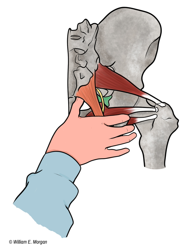
It carries sensory motor and autonomic fibers however an injury to the pudendal nerve causes sensory deficits more than motor. These are structures located near the genital rectal and gluteal buttock regions. After its formation the pudendal nerve descends and passes between the piriformis and ischiococcygeus muscles.
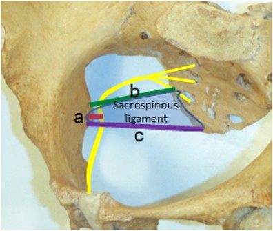
The pudendal nerve is the main nerve of the perineum. It courses between two muscles piriformis and coccygeus muscles. A rectal branch a perineal branch and a clitoralpenile branch.
The pudendal nerve is a sensory autonomic and motor nerve that carries signals to and from the genitals anal area and urethra. Anatomy of the pudendal nerve. The pudendal nerve is the main nerve that serves the perineum which is the area between the anus and the genitalia the scrotum in men and the vulva in women.
It arises from the ventral rami anterior divisions of the spinal nerves s2 s3 and s4. There are slight differences in the nerve branches for each person but typically there are three branches of the nerve on each side of the body. The condition known as pudendal neuralgia can cause both bladder and anal incontinence.
 Spotlight On A Pelvic Floor Diagnosis Pudendal Neuralgia
Pudendal Nerve Radiology Reference Article Radiopaedia Org
 Anatomy Of The Pudendal Nerve Health Organization For
 Pudendal Nerve Neuralgia Entrapment Springerlink
 Pudendal Nerve Block Atlas Of Pain Medicine Procedures
 Pudendal Nerve Anatomy Nervous Innervation Diagram Quizlet
![]() Sacral Plexus Anatomy Branches And Mnemonics Kenhub
Sacral Plexus Anatomy Branches And Mnemonics Kenhub
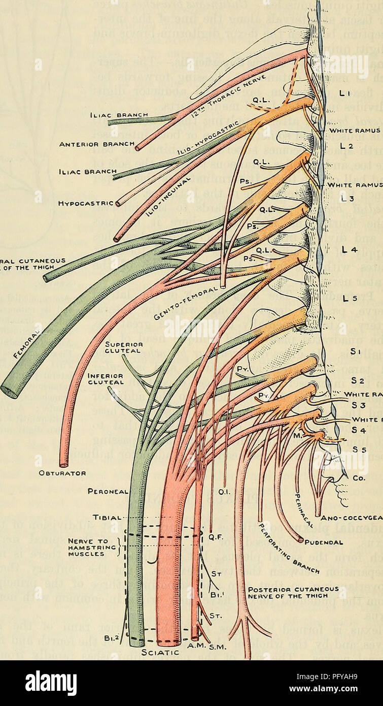 Cunningham S Text Book Of Anatomy Anatomy 736 The Neevous
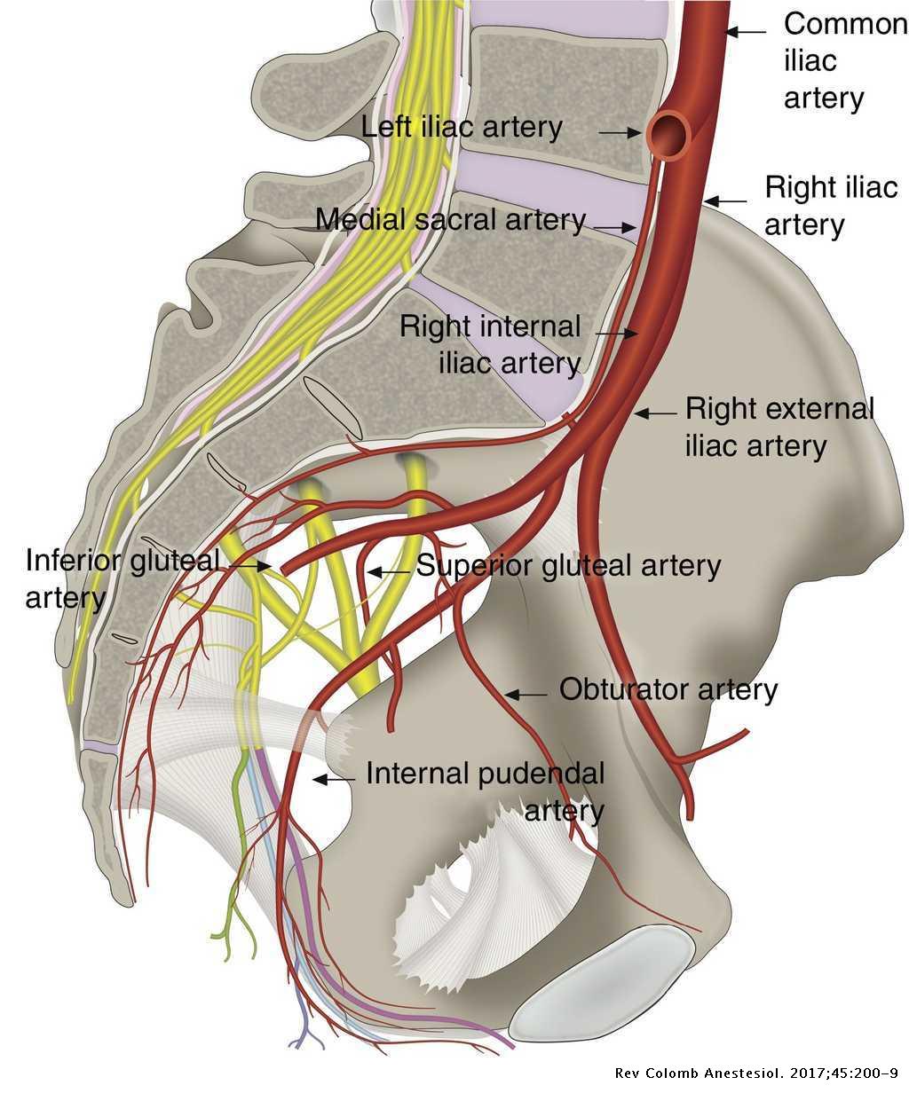 Regional Anesthesia Guided By Ultrasound In The Pudendal
 3t Magnetic Resonance Neurography Of Pudendal Nerve With
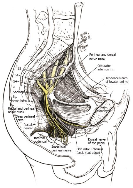
 Surgical Anatomy Of The Pudendal Nerve And Its Branches In
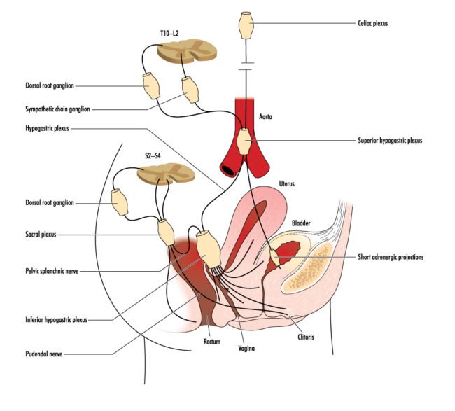
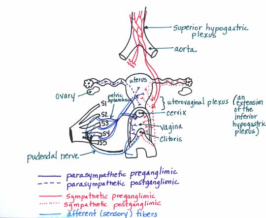 Module Autonomics Of The Pelvis
 Pudendal Nerve Wikipedia

 Figure 1 From Diagnosis And Treatment Of Pudendal Nerve
 Figure 3 From Anatomic Variations Of Pudendal Nerve Within
 Pudendal Neuralgia Ainsworth Institute Of Pain Management
 Clinical Pearl Pudendal Neuralgia Cyclist S Syndrome
 Pudendal Neuralgia Physiopedia
 Pudendal Nerve Entrapment Springerlink
 Pudendal Nerve An Overview Sciencedirect Topics
 Anal Canal Anatomy Gross Anatomy Tissue Nerves And
 Sacral Plexus Nerve Plexus Lumbar Plexus Anatomy Pudendal
 Regional Anesthesia Guided By Ultrasound In The Pudendal
 Plexus And The Pudendal Nerve Buscar Con Google Plexus
 Pudendal Nerve Blockade Springerlink
 Pudendal Neuralgia And Complex Pelvic Pain An Interview
 Anatomy Of The Pudendal Nerve Health Organization For
 The Pudendal Nerve Anatomical Course Functions
 3d Of Pudendal Nerve Www Dellon Com
![]() Sacral Plexus Anatomy Branches And Mnemonics Kenhub
Sacral Plexus Anatomy Branches And Mnemonics Kenhub
Belum ada Komentar untuk "Pudendal Nerve Anatomy"
Posting Komentar