Anatomy Of The Heel Of The Foot
The main ligaments of the foot are. The deep fibular peroneal nerve runs through extensor digitorum longus and down the interosseous membrane.
 Foot And Ankle Anatomical Chart
The foot is divided into three sections the forefoot the midfoot and the hindfoot.
Anatomy of the heel of the foot. The heel is the portion of the human body that lies at the bottom rear part of each foot. The ligament which runs along the sole of the foot from the heel to the toes forms the arch. The talus ankle bone is the second largest of the tarsal bones and sits atop the calcaneus.
The calcaneus heel bone is the largest bone in the foot. Plantar fascia the longest ligament of the foot. The heel bone is the largest bone in the foot.
Then it crosses the tibia and enters the dorsum of the foot. It is the is the strongest and largest tendinous structure in the body. The rear half of the heel bone is known as the tuber calcanei.
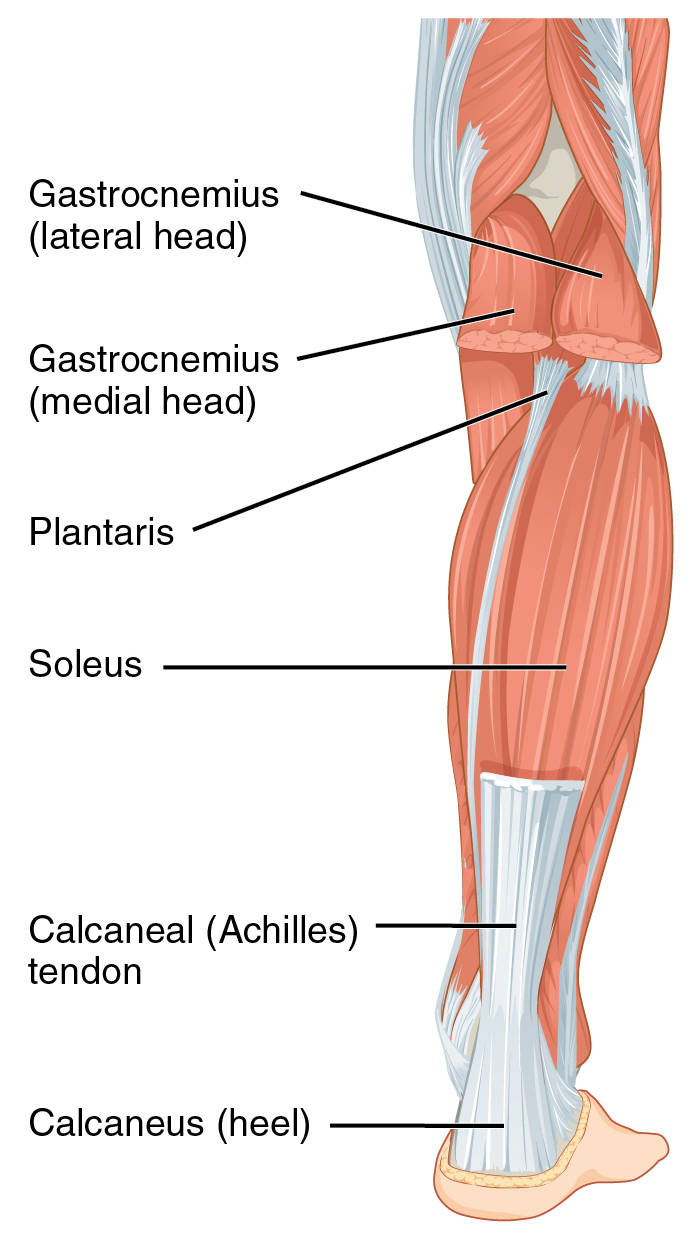
The foot consists of thirty three bones twenty six joints and over a hundred muscles ligaments and tendons. The achilles tendon makes it possible to run jump climb stairs and stand on your. The most notable tendon of the foot is the achilles tendon which runs from the calf muscle to the heel.
The heel bone is designed to be the first contact the foot has with the ground. It innervates the muscles in the anterior compartment of the leg and the dorsum of the foot. These all work together to bear weight allow movement and provide a stable base for us to stand and move on.
The achilles tendon inserts into the back of the heel bone calcaneus and a very strong ligament along the bottom of the foot attaches to the bottom of the heel bone the plantar fascia. Muscles tendons and ligaments run along the surfaces of the feet allowing the complex movements needed for motion and balance. Foot and ankle anatomy is quite complex.
By stretching and contracting the plantar fascia helps us balance and gives the foot strength for walking. It also supplies a small region of skin between the first big and second toes. The calcaneus joins with the talus and cuboid.
Foot and ankle anatomy. The calcaneus heel bone is the largest of the tarsal bones. The anatomy of heel pain.
It transmits the weight of the body to the ground and acts as a lever for the muscles of the calf. Heel in anatomy back part of the human foot below the ankle and behind the arch and the corresponding part of the foot in other mammals that walk with their heels touching the ground such as the raccoon and the bear. The anatomy of the foot the foot contains a lot of moving parts 26 bones 33 joints and over 100 ligaments.
It corresponds to the point of the hock of hoofed mammals and those that walk on their toes eg horse dog cat.
 Achilles Tendon Human Anatomy Picture Definition
 Duke Anatomy Lab 2 Pre Lab Exercise
Heel Spur Syndrome Foot Ankle Doctors Inc
 Foot Anatomy And Function प द Pada Elliots World
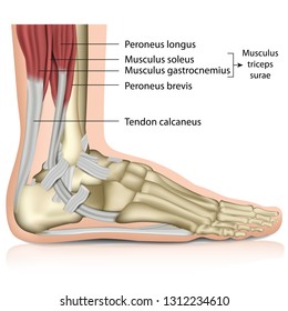 1000 Calcaneus Stock Images Photos Vectors Shutterstock
 Foot Pain Diagnosis Achilles Tendinitis Causes Home
 Lower Legs And Feet Running Anatomy Sports Anatomy
Foot Pain And Heel Pain Body Active
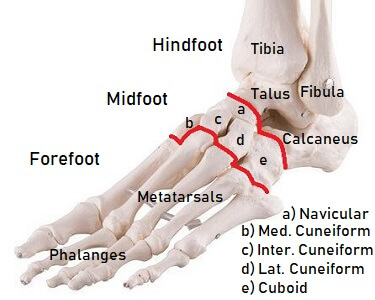 Foot Bones Anatomy Injuries Foot Pain Explored
 Foot Ankle Anatomy Pictures Function Treatment Sprain Pain
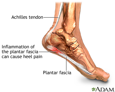 Plantar Fasciitis Medlineplus Medical Encyclopedia
 The Foot And Ankle Practical Office Orthopedics
Foot Heel Pain Identifying Plantar Fasciitis
Anatomy Heal The Heel Pain Plantar Fasciitis
 Sky Soles Foot Anatomy And High Heels Galley Gossip Blog
 Ankle And Foot Exam Stanford Medicine 25 Stanford Medicine
 Duke Anatomy Lab 2 Pre Lab Exercise
 Pin On Foot And Ankle Health Tips
 Is Your Foot Pain Caused By A Spine Problem
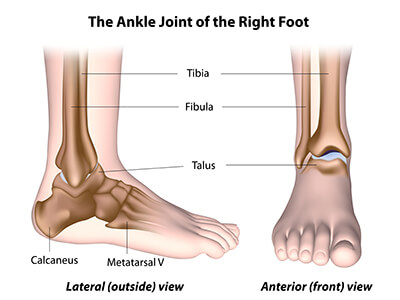 Ankle Cartilage Preservation Towson Orthopaedic Associates
Flexor Hallucis Longus Dysfunction Academy Of Clinical Massage
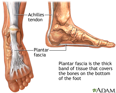 Plantar Fasciitis Medlineplus Medical Encyclopedia
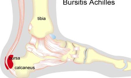 Foot Achilles Arkiv Sportnetdoc
Heel Pain Foot Specialists Of Shreveport Bossier
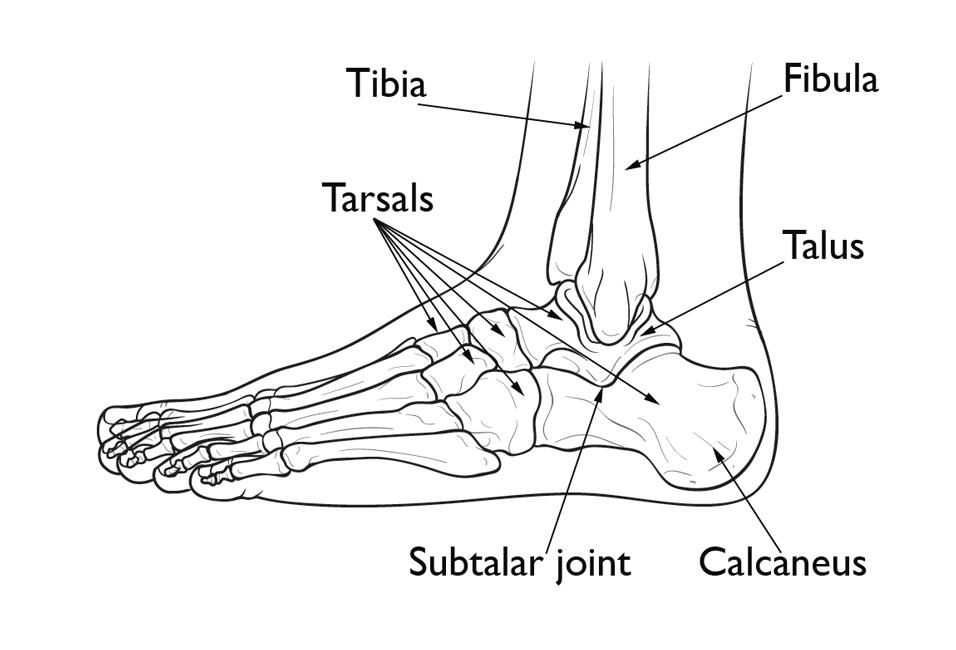 Calcaneus Heel Bone Fractures Orthoinfo Aaos
Belum ada Komentar untuk "Anatomy Of The Heel Of The Foot"
Posting Komentar