Urinary Bladder Anatomy
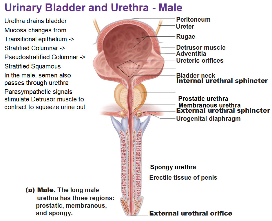
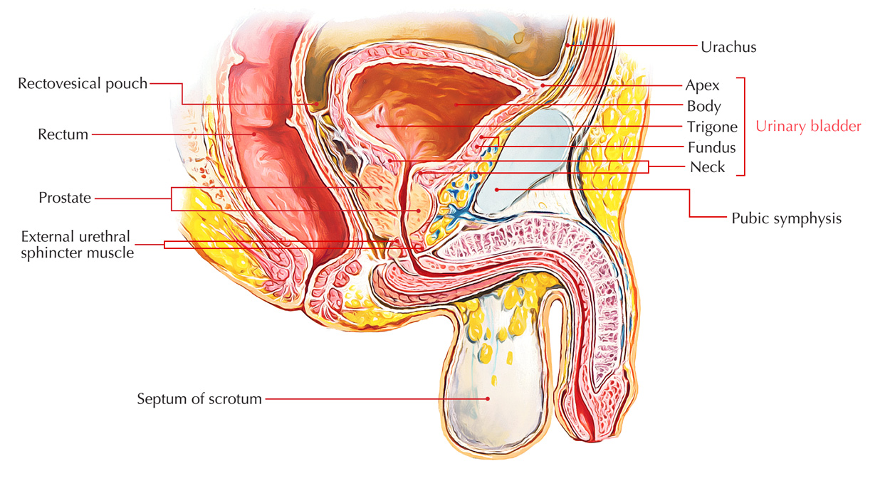
Urine leaves the bladder via the urethra a single muscular tube ending in an opening the urinary meatus. In males the vas deferens sperm carrying tubes empty into the urethra.
 The Anatomy Of The Urinary Bladder Biology Forums Gallery
It has a muscular elastic wall which allows it to expand and distend when filled.

Urinary bladder anatomy. The appearance of the bladder varies depending on the amount of urine stored. In females the urethra is separate from the genital tract. The urinary bladder is hollow and somewhat pear shaped in appearance.
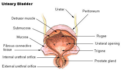
Surrounding the mucosal layer is the submucosa a layer of connective tissue with blood vessels. The innermost layer of the bladder is the mucosa layer that lines the hollow lumen. The urinary bladder is a hollow muscular organ.
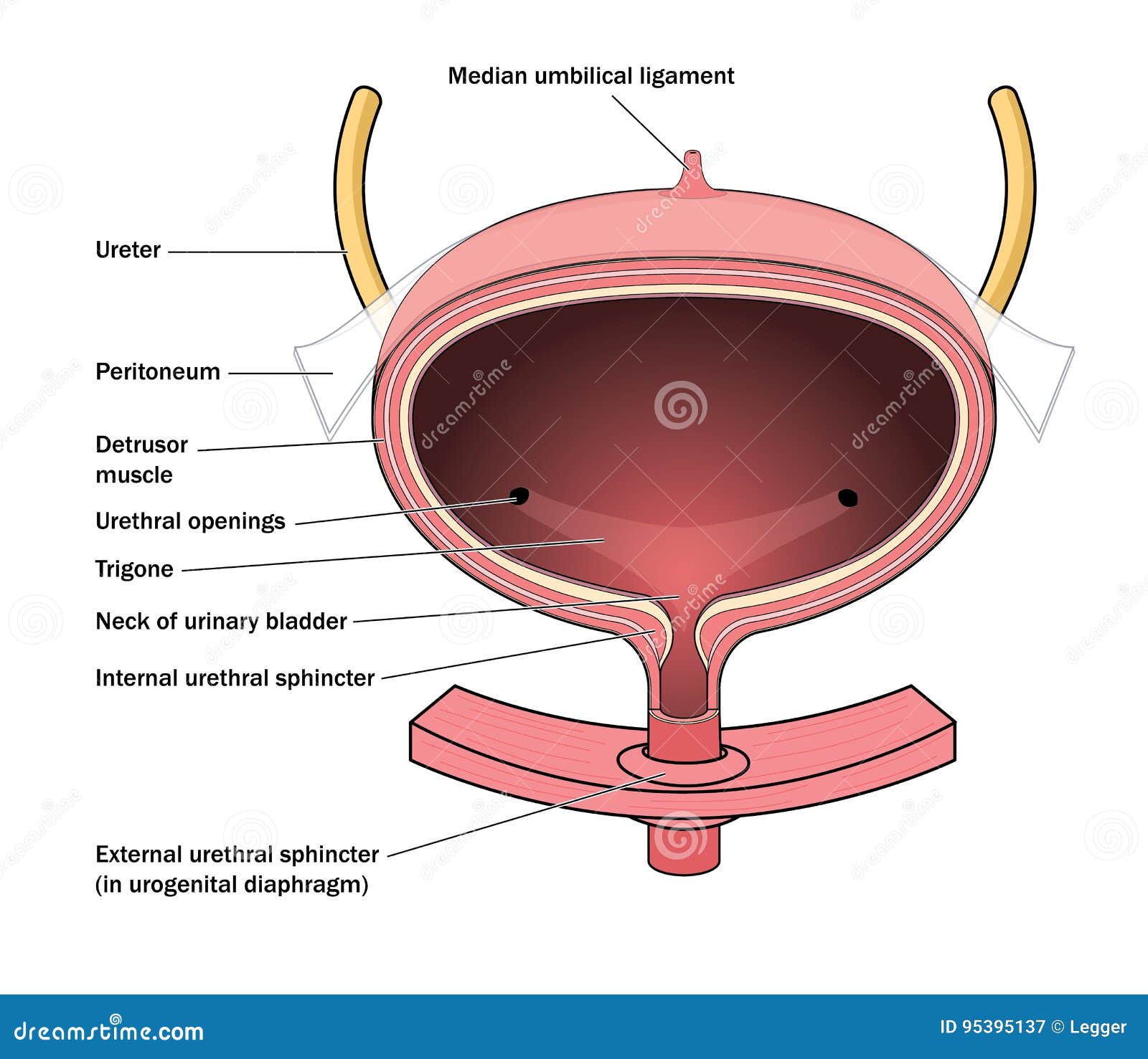
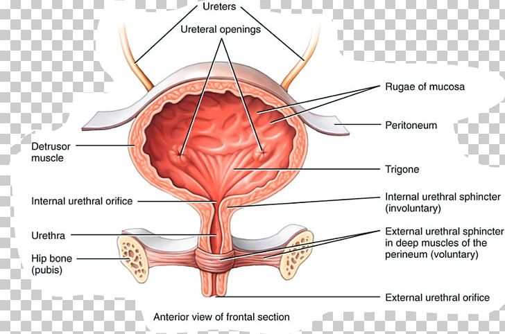
The interior of the bladder are the trigone of the bladder. The bladder has a volume capacity of 400500 ml and is. In placental mammals a special duct the urethra leads from the urinary bladder to the exterior.
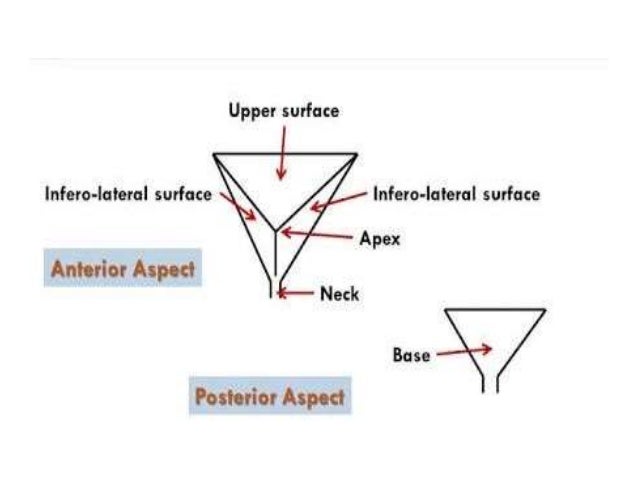
When empty the bladder is about the size and shape of a pear. The urinary bladder usually just called the bladder is a major part of the bodys urinary system1 3 it is a hollow organ that is made mostly out of muscle. The urinary bladder shape of the bladder.
The sphincter of the urinary bladder consists of smooth. It temporarily stores urine conveyed by the ureters from the kidneys until the body is ready to expel it through the urethra. The visceral muscles of the muscularis layer.
The urinary bladder is made of several distinct tissue layers. Interior of the bladder. Sphincter of the urinary bladder.
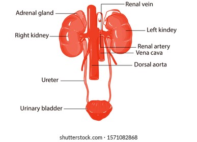
The vasculature of the bladder is primarily derived from the internal iliac vessels. The bladders job in the urinary system is to store the urine produced by your body until it is released from the body when you urinate. Urine collects in the bladder fed from the two ureters that connect the bladder with the kidneys.
The urinary bladder is a muscular sac in the pelvis just above and behind the pubic bone. In humans the bladder is a hollow muscular organ situated at the base of the pelvis. The superolateral aspect of the bladder drains into the external.
Urine is received into the body of the. Gross anatomy of the bladder surface anatomy of the bladder. Urinary bladder the urinary bladder is an organ that serves to collect urine to be voided through urination after the urine is filtered through the kidneys where the necessary ions are reabsorbed if physiologically needed through feedback mechanisms found throughout the body and in the nephrons of the kidneys such as the macula densa.
It fulfills the excretory function of the more primitive cloaca.
 Urinary Bladder Function Blood Supply And Innervation Human Anatomy Kenhub
 Royalty Free Urinary Bladder Stock Images Photos Vectors

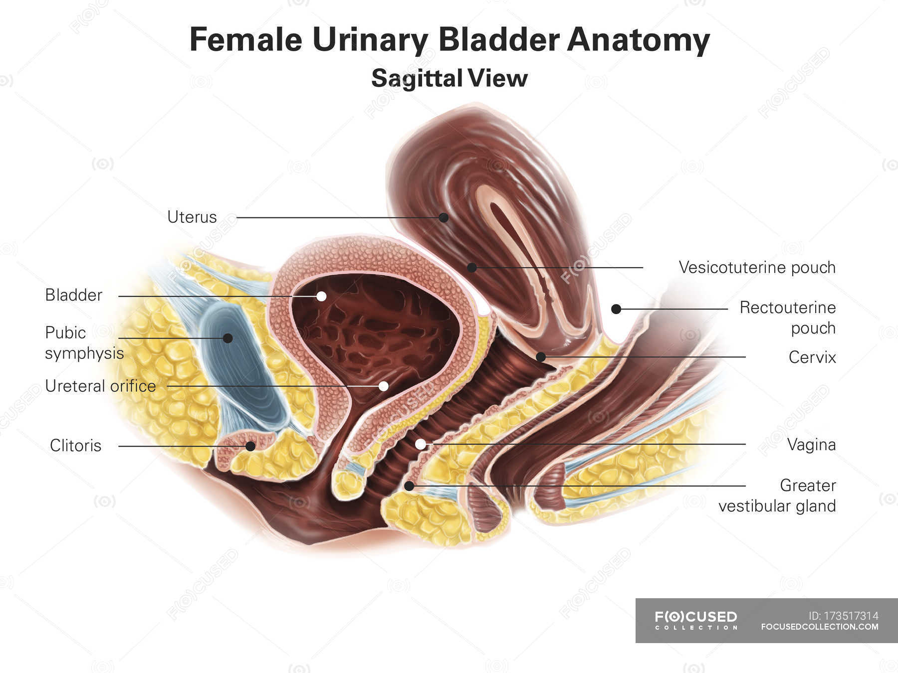 Female Urinary Bladder White Background Digitally
 Seer Training Layers Of The Bladder Wall
 Anatomy Of The Urinary Bladder Stock Vector Illustration
 The Urinary Bladder Is A Muscular Sac Composed Of 3 Layers
 Anatomy Of Urinary Bladder
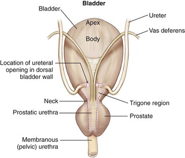
 Male Urinary Bladder Diagram Quizlet
 Dictionary Normal Urinary Bladder The Human Protein Atlas
 Urinary Bladder Anatomy Qa

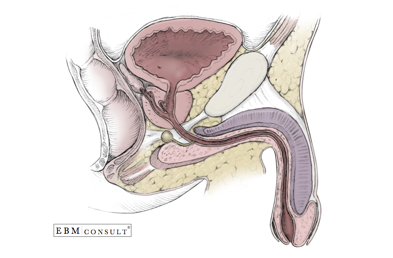 Male Genitourinary Anatomy Bladder Prostate Penis In
 Seer Training Urinary Bladder
 The Urinary System Ureter And Urinary Bladder
 American Society Of Clinical Oncology Endorses Eau Guideline
 Anatomy Female Urinary Bladder Wall Decal
 Urinary System Anatomy Urinary Bladder Examination Normal And
 Anatomy Female Urinary Bladder Wall Mural
Bladder Urethra Anatomy Renal Medbullets Step 1
 Bladder Veterian Key
 Easy Notes On Urinary Bladder Learn In Just 4 Minutes
 Stones In The Urinary Bladder And Ureter Human Anatomy Illustration
 Urinary Bladder Anatomy Excretory System Urine Autonomic
 Urinary Bladder Wikipedia
Belum ada Komentar untuk "Urinary Bladder Anatomy"
Posting Komentar