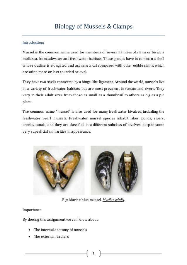
Mussel Anatomy
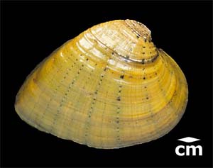
The umbo of the zebra mussel is the genesis growing point of its shells which produces the growth rings visible on the the exterior portion of the shell. The middle layer of mussel shell which is composed of column shaped crystals of calcium carbonate caco3.
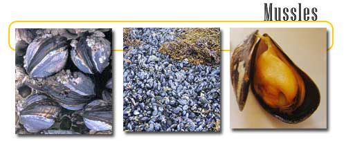
 Bouchot Mussels Green Lip Mussels Gabe The Fish Babe
Owl pellets carolina provides owl pellet products that are heat sterilized and easy to use for students of all ages.
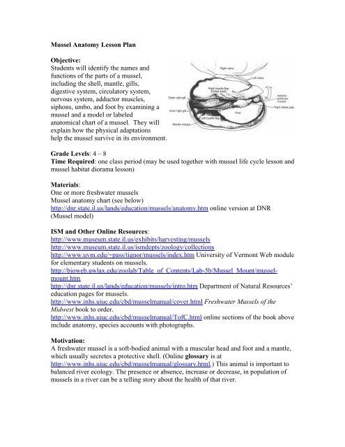
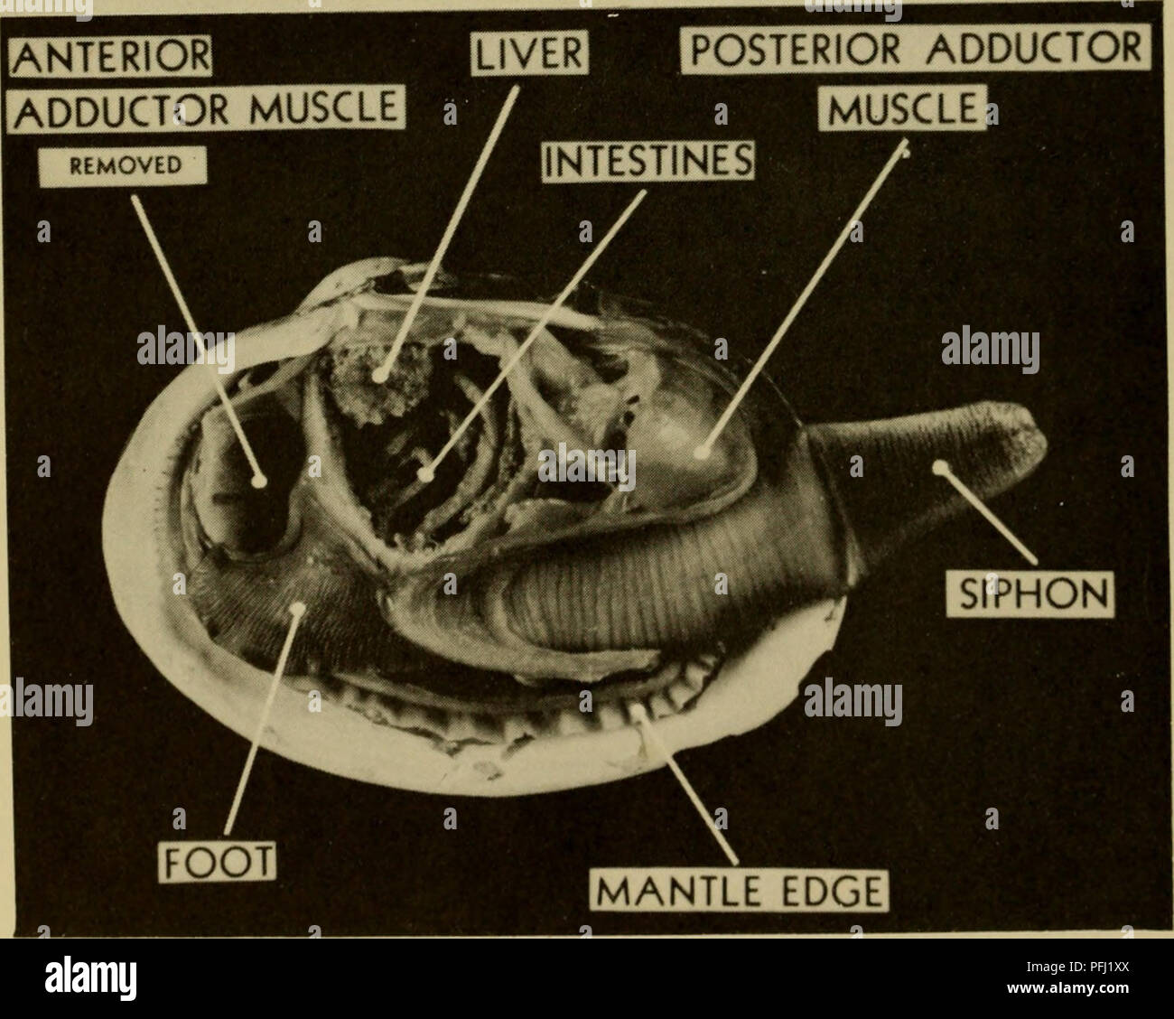
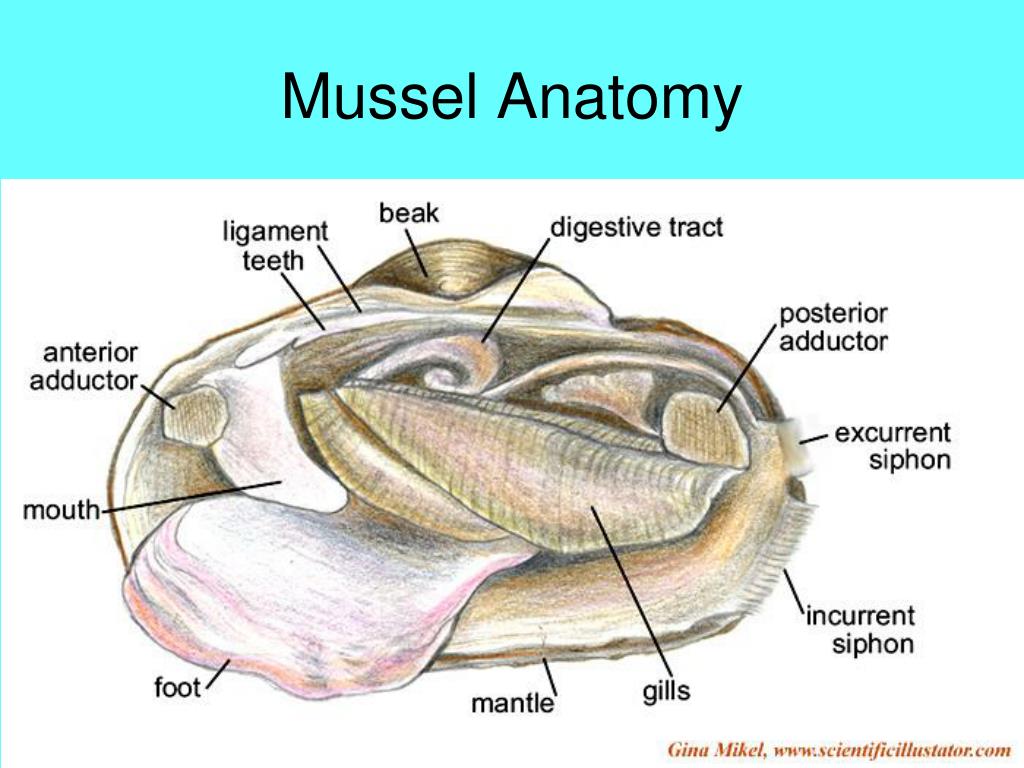
Mussel anatomy. Marine mussels are usually found clumping together on wave washed rocks each attached to the rock by its byssus. Pseudocardinal teeth noun triangular often serrated structures hinge teeth located near the anterior dorsal margin of some valves. Sheetlike prominently striated structures in the dorso posterior region a pair on each side of the body dorsal margin is attached but not ventral microscopic cilia of these more water and suspended food along toward the mouth.
Perna canaliculus inhibits the 5 lipoxygenase pathway which leads to the formation of leukotrienes. The shells of different species vary in size shape thickness and color. We think this is the most useful anatomy picture that you need.
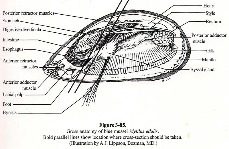
For more anatomy content please follow us and visit our website. This is a great way to learn basic anatomy. The breakdown of food particles is facilitated by these enzymes and the mixing of the stomach contents by the rotation of the crystalline style against the gastric shield.
Anatomy of freshwater mussels. These rings are slightly visible in the picture of the zebra mussel below. At low tide mussels in the middle of a clump will undergo less water loss because of water capture by the other mussels.
Shells also vary in the presence or absence of sculpturing. It differs from other mussel species in that it has dark browngreen shells with green lips around the edges and has only one adductor muscle. In all bivalves the style is a gelatinous rod like body that contains starch digesting enzymes and is continually being used up and renewed.
We hope this picture mussel anatomy diagram can help you study and research. Mussel anatomy and physiology. It is also one of the largest mussel species reaching 240 mm in length.
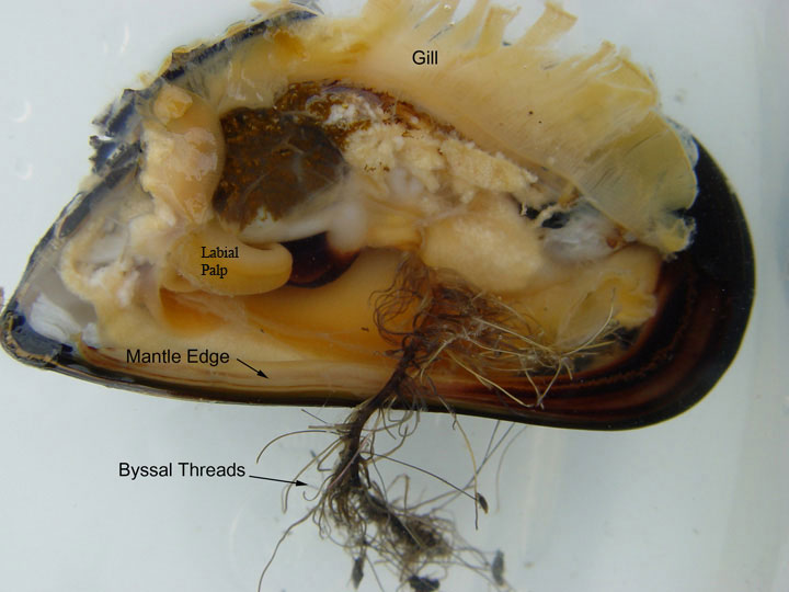
The clumping habit helps hold the mussels firm against the force of the waves. Shell anatomy of the zebra mussel. The shells each mussel has two shells one left valve and one right valve that protect the soft bodied animal from predators.

 Anatomy Of The Mussel Doi 10 1371 Journal Pone 0008875 G001
 File Diagram Freshwater Mussels Jpg Wikimedia Commons
 Mussel Anatomy Diagram Quizlet
Influence Of Wave Action On Mytilus Californianus Mussel Beds
 Amazon Com Mussels Anatomy Habitat And Environmental
 The Anatomy Of The Freshwater Mussel Dvd Amazon Com
 Dangerous Marine Animals Marine Animals Foot Byssus Gills
 3d Mussel Mytilus Edulis Anatomy
 Mussel Anatomy Clipart Etc
 Chm101h1 Lecture 4 Animals Ii Mussel Dissection 2018 Oneclass
 Pin By Bob On Freshwater Molluscs Primarily Unionids
Anatomical Features Of A Freshwater Mussel With The Right
 External A And Internal B Shell Morphology Of Charru

 Biology Of Mussels Camps
 I Don T Understand Mussels Anatomy But What S This Long
 Mussel Anatomy Left Shell Diagram Quizlet
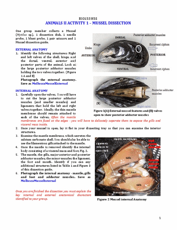
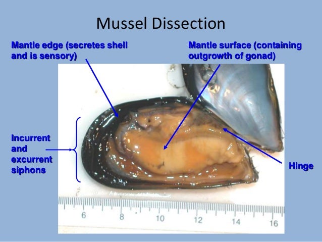 Mussel Dissection
 Shellfish Roger Williams University
 Mid Intertidal Zone
 Education
 Ppt Phylum Mollusca Powerpoint Presentation Free Download
Mussel Anatomy Lesson Plan Illinois State Museum
 Bivalve Anatomy Shells Anatomy Mussels
Belum ada Komentar untuk "Mussel Anatomy"
Posting Komentar