Mri Prostate Anatomy
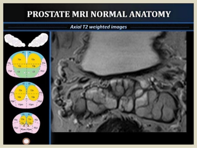
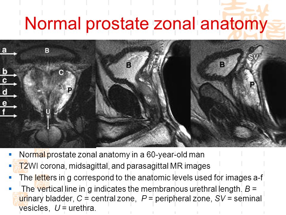
There is no ionizing radiation x rays used to create any mri image. The peripheral zone showed higher signal intensity than either the central or transition zone and was discerned in the coronal sagittal and transverse planes.
 Introduction To Prostate Anatomy On Mri Mrionline 8
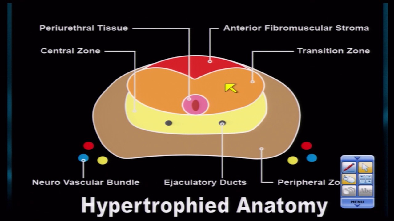
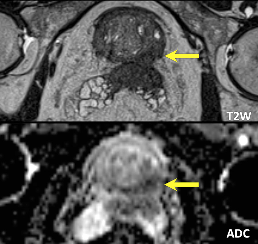
Small bph nodule at r base at the centralperipheral zone border benign prostate hyperplasia bph typically arises in the transition zone distorts zonal anatomy and can get very large over 200 ml.

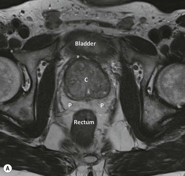
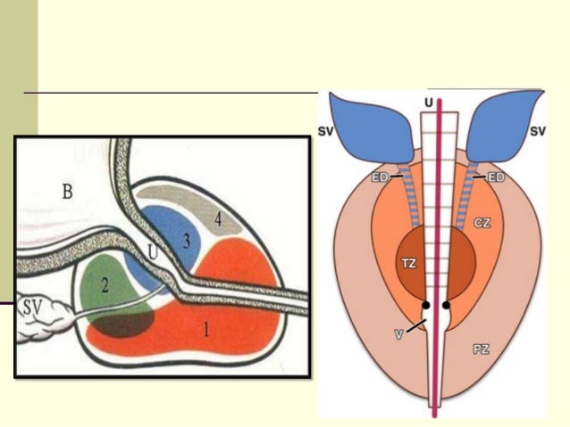
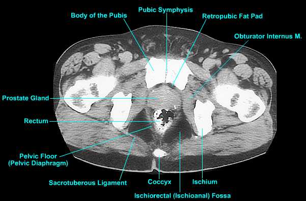
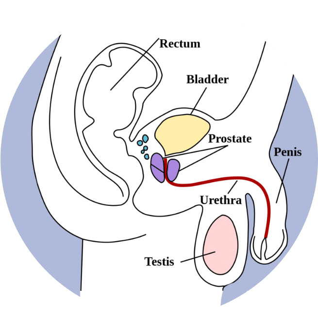
Mri prostate anatomy. Prostate anatomy mri radiology assistant. As health systems around the world adopt mri as the preferred method to assess diagnose and monitor the progression of prostate cancer the demand has increased for detailed educational material for radiologists who read prostate mrs. The prostate gland is a small soft structure about the size and shape of a walnut which lies deep in the pelvis between the bladder and the penis and in front of the rectum back passage.
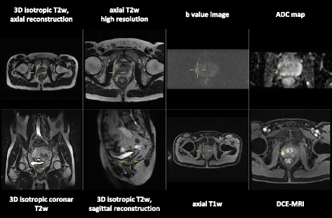
In prostate cancer patients mri may be used to examine the prostate and nearby lymph nodes to distinguish between benign noncancerous. Multiparametric mri is a combination of t2 weighted diffusion and dynamic contrast enhanced imaging and is an accurate tool in the detection of clinically significant prostate cancer. Using computer aided detection cad this exam can pinpoint specific areas within the prostate gland that are suspicious and may require further evaluation.
Mri for diagnosis and treatment of prostate cancer duration. The majority of cancers arise from the peripheral zone pz. Mri of the prostate has become increasingly popular with the use of multiparametric mri and the pi rads classification.
Instead mri uses a large magnet radio waves and a computer to produce these images. Evaluating sequential sections the peripheral central and transition zones could be differentiated. An mri is a test that produces very clear pictures of the human body without the use of x rays.
Prostate hyperplasia bph less commonly occurs outside of the transition zone. A magnetic resonance imaging mri scanner uses strong magnetic fields to create an image or picture of the prostate and surrounding tissues. Prostate mri captures detailed images of the prostate to evaluate for prostate cancer or disease.
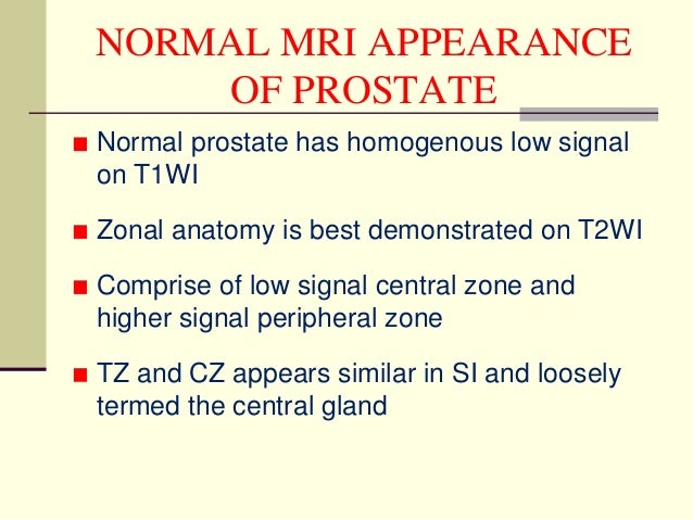
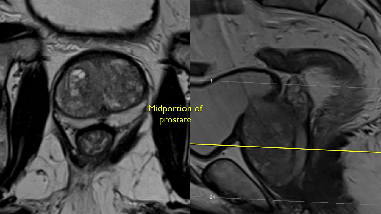
Prostate mri case review a continued look at anatomy 30 duration. Mri imaging is helpful in differentiation the prostatic zonal anatomy best demonstrated on t2wi. Mr imaging of the prostate gland.
Special techniques are used to improve the early detection of prostate cancer such as dwi dynamic contrast enhanced mri and mr spectroscopy. An mri study of the prostate incorporates a powerful magnet system radio waves and a computer to create very detailed images of the prostate gland and surrounding anatomy.
 Prostate Radiology Key

 Prostate Mri Based On Pi Rads Version 2 How We Review And
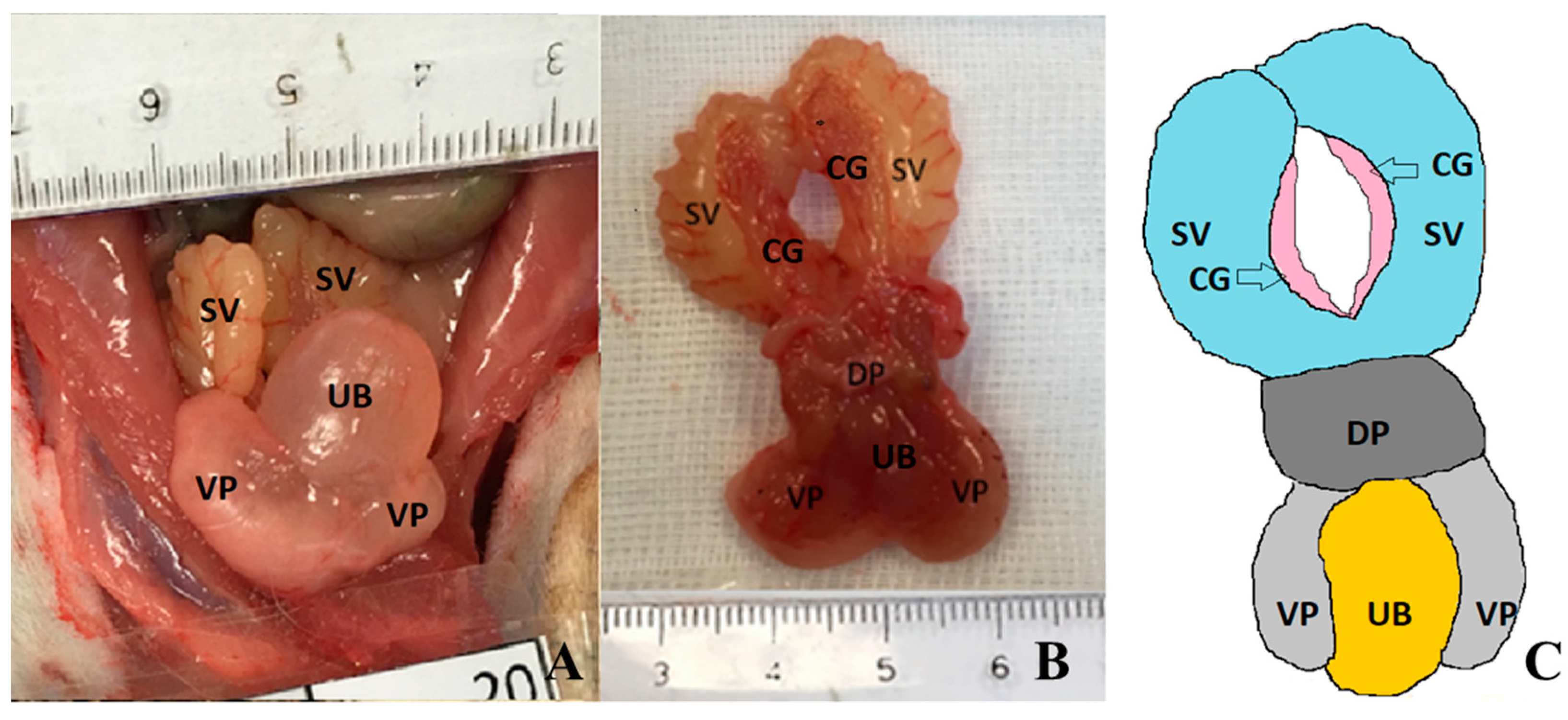 Diagnostics Free Full Text Anatomy And Imaging Of Rat
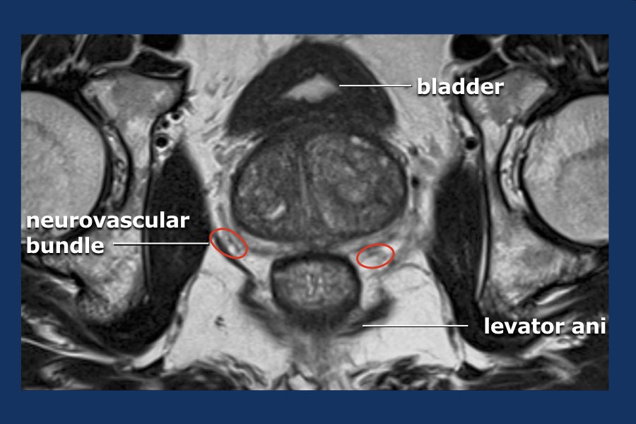 The Radiology Assistant Prostate Cancer Pi Rads V2
 Mri Prostate
 Mri Male Pelvis Anatomy Free Male Pelvis Sagittal Anatomy
 Prostate Anatomy Mri
 Mri Prostate
 Mri Prostate
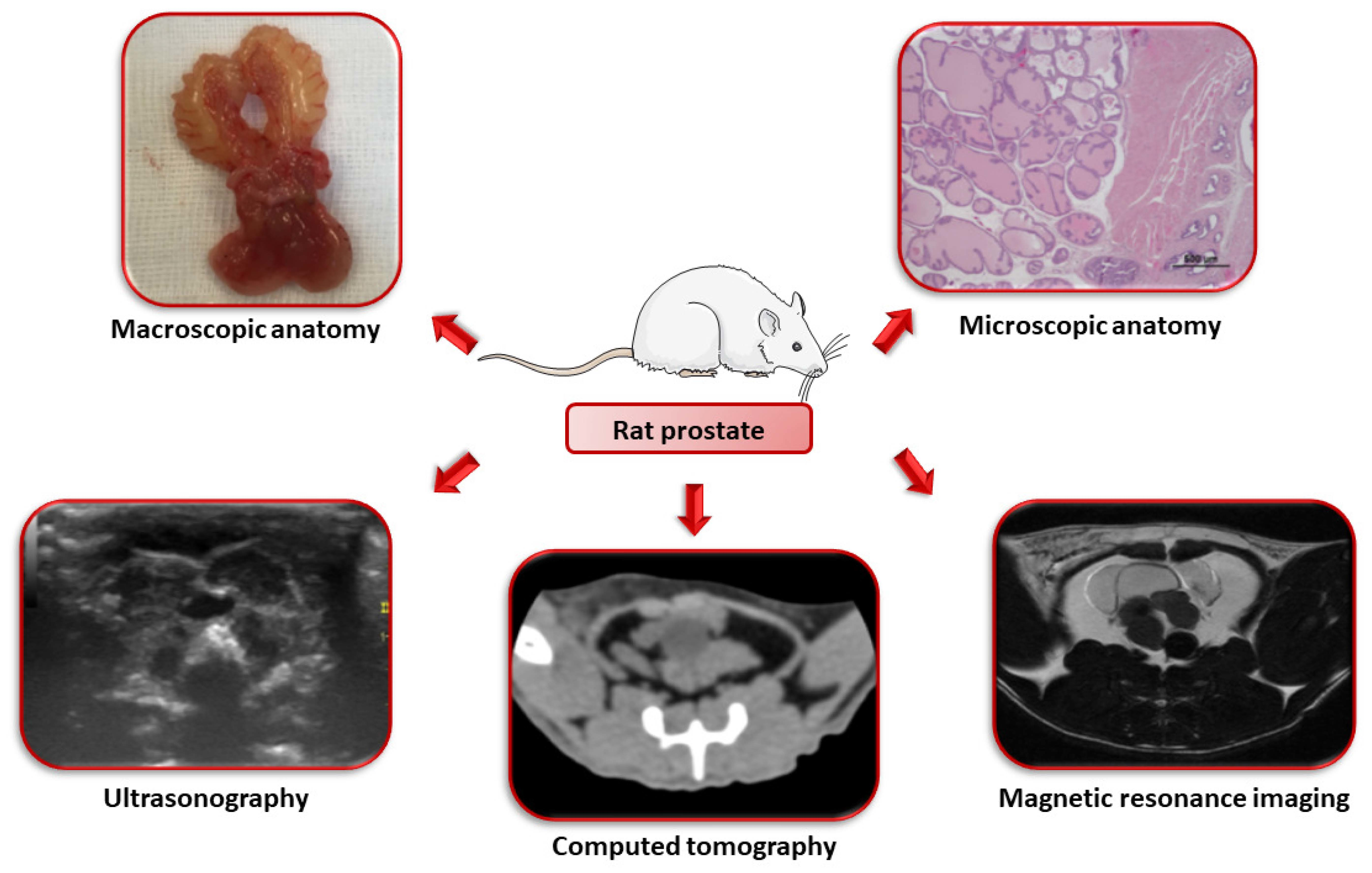 Diagnostics Free Full Text Anatomy And Imaging Of Rat
 Normal Prostate Mri Radiology Case Radiopaedia Org
 Multiparametric Mr Imaging In Diagnosis Of Chronic
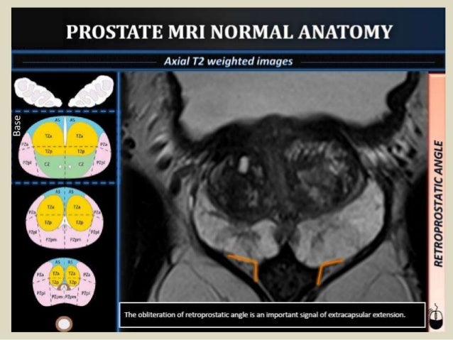 Presentation1 Mri Imaging Of The Prostate
 Prostate Mri Case Review A Continued Look At Anatomy 30
Multiparametric 3t Mr Imaging Of The Prostate Acquisition

 The Radiology Assistant Prostate Cancer Pi Rads V2
 Normal Prostate Anatomy On Magnetic Resonance Imaging T1 And

 Presentation1 Mri Imaging Of The Prostate
 The Radiology Assistant Prostate Cancer Pi Rads V2

 Genitourinary Imaging Prostate Ppt Download
 Full Body Ezra Detect Cancer Early Using Mri And Ai
 Clinical Background And Needs Cobra

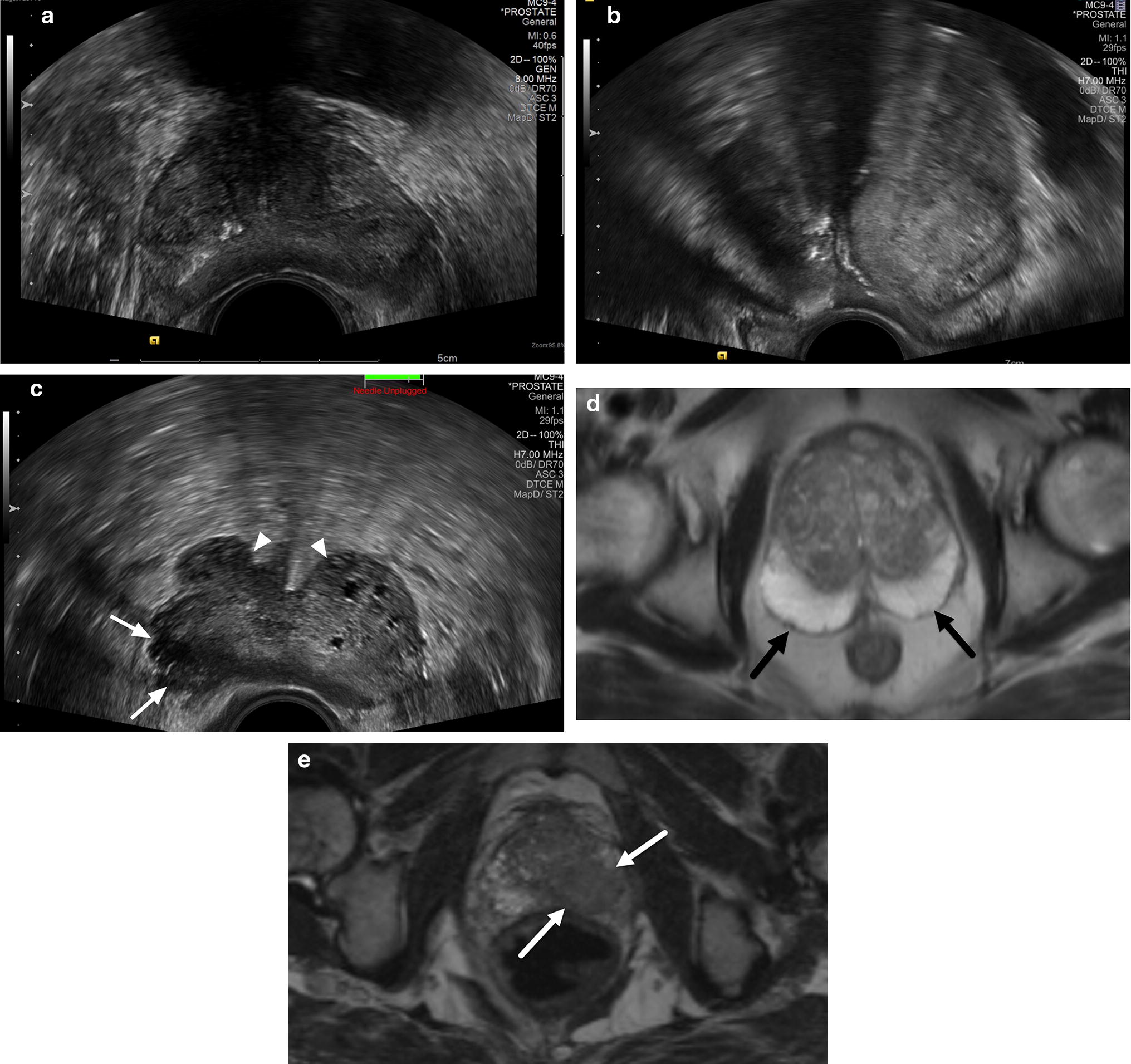 Prostate Cancer Detection And Diagnosis Role Of Ultrasound
 The Male Pelvis Mr Anatomy Atlas Of The Prostate Bladder
Belum ada Komentar untuk "Mri Prostate Anatomy"
Posting Komentar