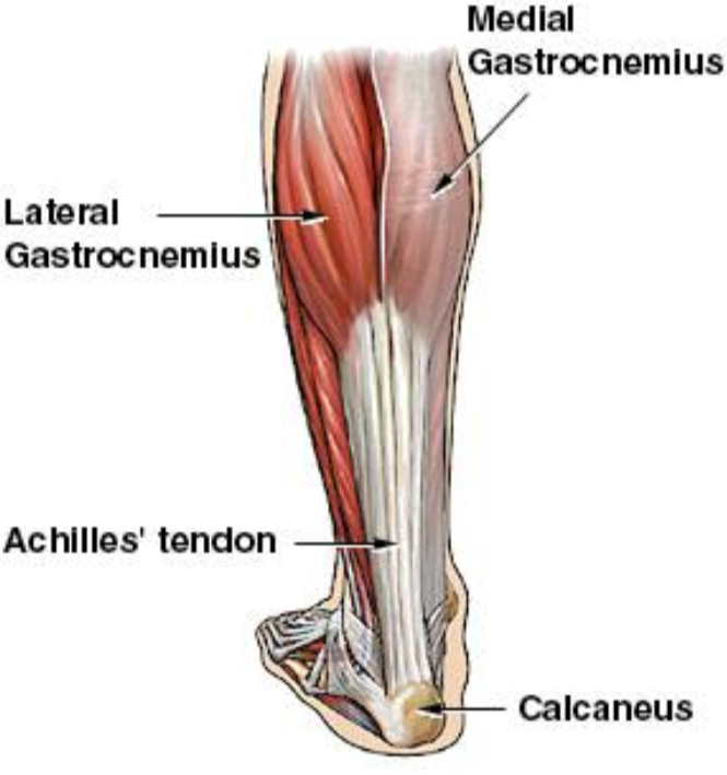
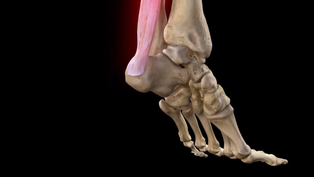
Achilles Tendon Anatomy
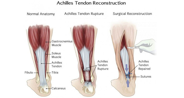
The achilles tendon is a tough band of fibrous tissue that connects the calf muscles to the heel bone calcaneus. It is the largest and strongest tendon in the human body and is capable of supporting tensional forces produced by movement of the lower limb.
 Achilles Tendon Rupture Diagnosis Causes Treatment
The anatomy of the tendon provides for both elasticity recoil and shock absorbance in the foot.

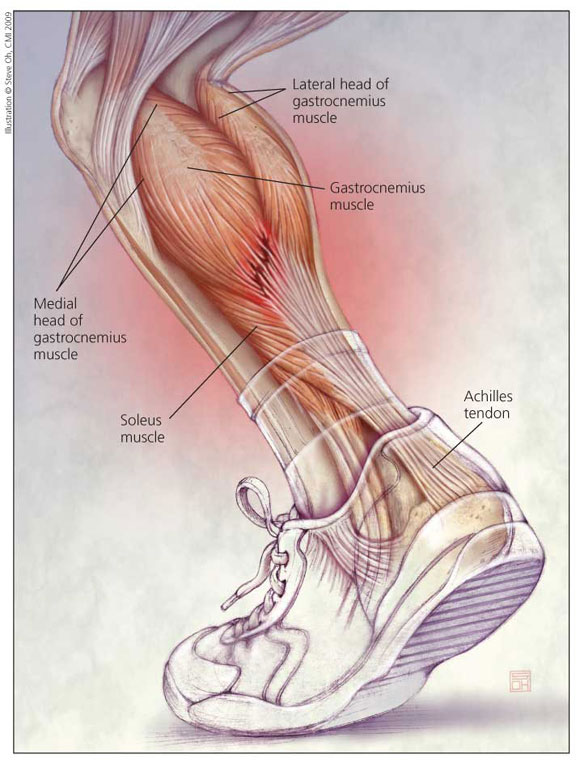
Achilles tendon anatomy. The achilles tendon is also called the calcaneal tendon. The achilles tendon at is the thickest and strongest tendon in the human body. Its origin lies close to the middle of the calf and fuses with the gastrocnemius muscle proximally.
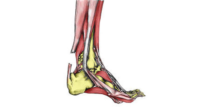
Anatomy of the achilles tendon the achilles tendon also known as the calcaneal tendon is a white fibrous cord located at the back of the ankle. The blood supply to the achilles tendon forms a network of arteries within the paratenon covering the tendon surface 9. The gastrocnemius is a fusiform muscle formed by two heads medial and lateral each separately crossing the knee joint.
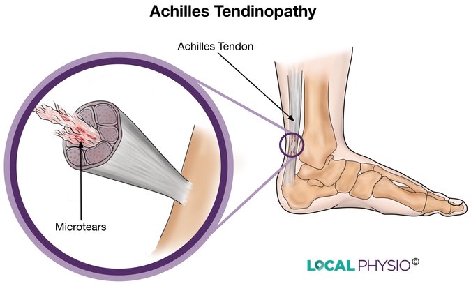
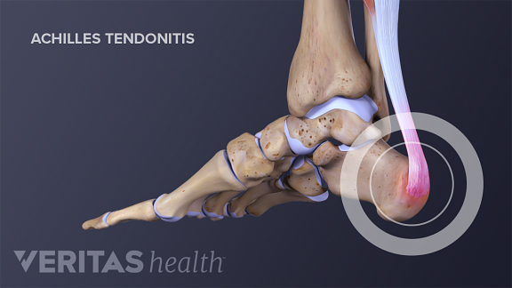
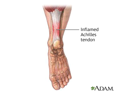
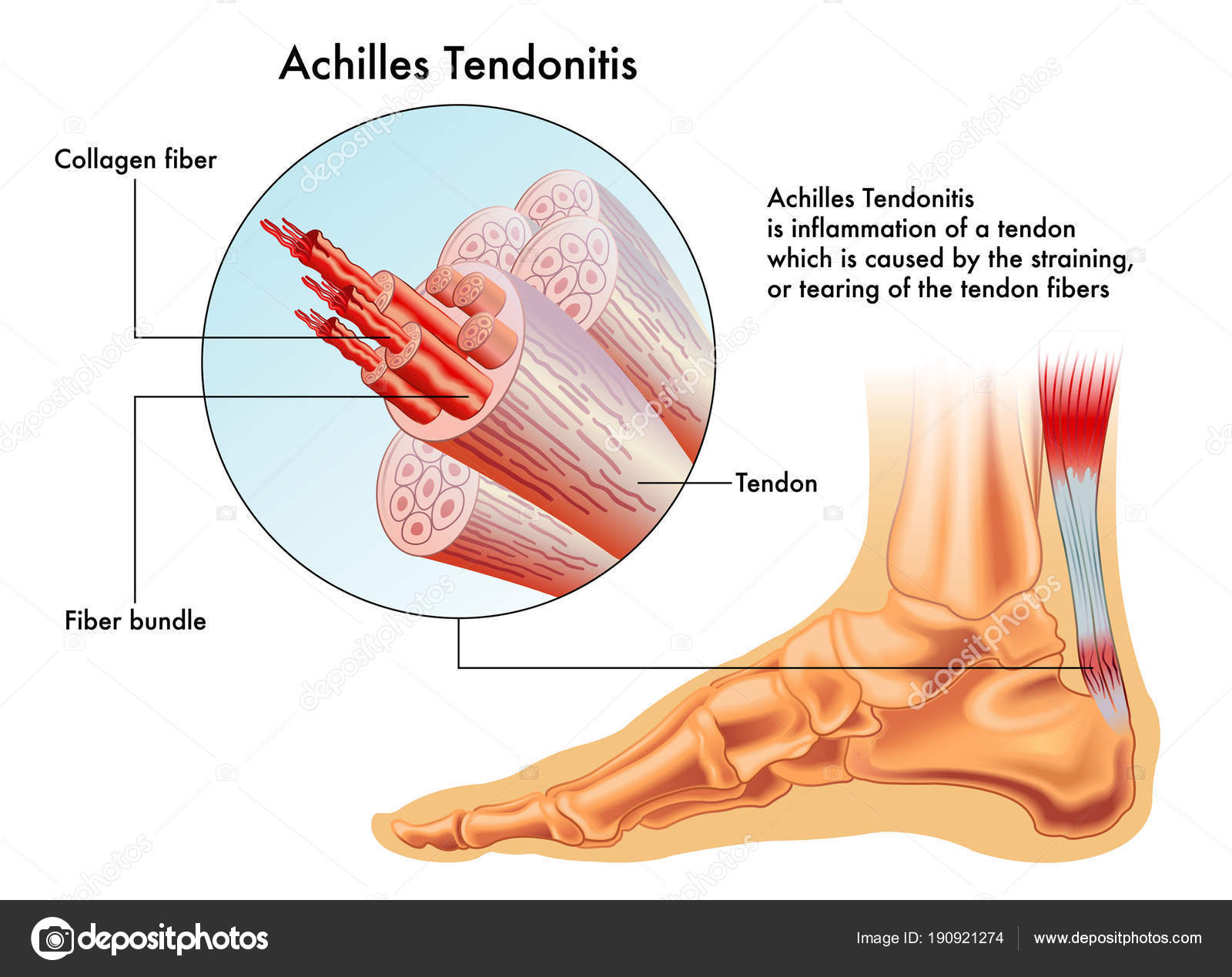
The majority of the achilles tendon is supplied by branches of the posterior tibial artery which are located medial to the tendon and supply the proximal and distal portions of the tendon 9. The achilles tendon is one of the most robust tendons in the body and for good reason. The achilles tendon is susceptible to damage with repetitive use or overload.
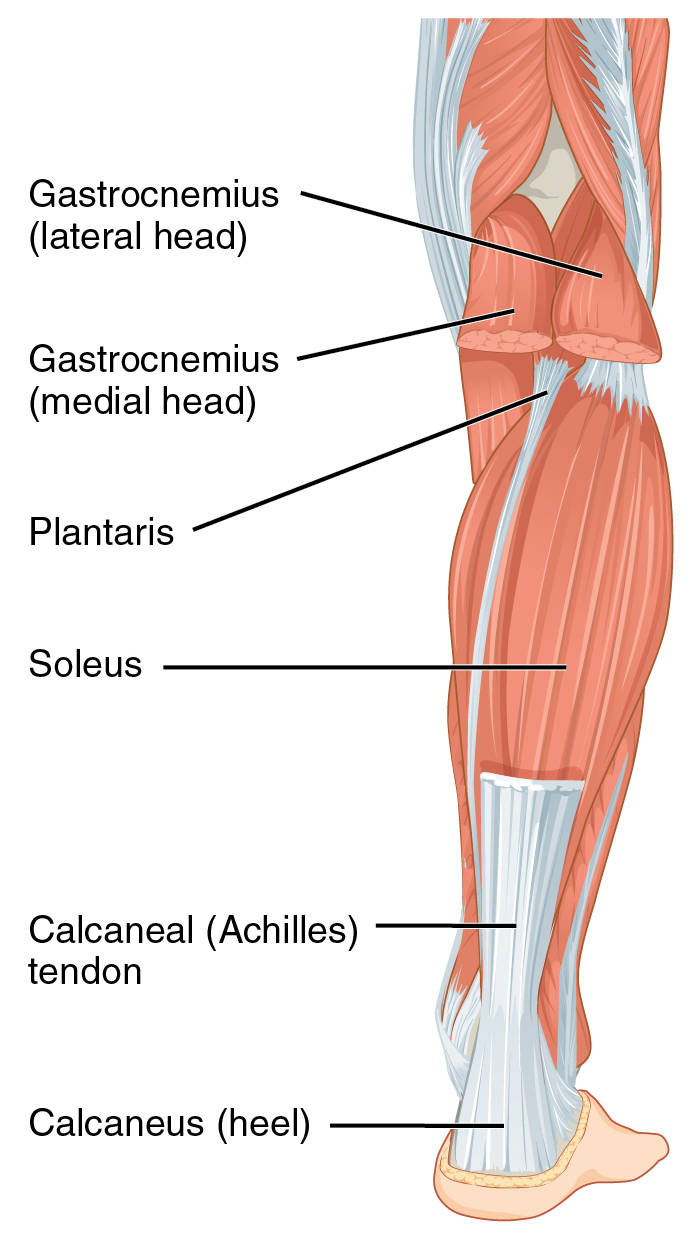
Three relatively large and extremely strong muscles in the calf the gastrocnemius soleus and plantaris all attach to the back of the heel bone calcaneus via the achilles and the forces they generate during running and jumping are immense among the biggest in the body. Learn about the anatomy and vulernability to injury of the achilles tendon. Achilles tendon strong tendon at the back of the heel that connects the calf muscles to the heel.
Essential in the flexion of the subtalar joint also known as the talocalcaneal joint in the ankle which exists between the calcaneus heel bone and the talus bone. The tendon is formed from the gastrocnemius and soleus muscles. It is named after the ancient greek mythological figure achilles.
Anatomy and importance of the achilles tendon the achilles tendon tendo calcaneus or tendo achillis is the thickest and strongest tendon in the human body. It is the tendinous extension of the three headed calf muscle consisting of soleus and the two headed gastrocnemius.
 Achilles Tendon Wikipedia
 What Do We Know About Achilles Tendinopathy Onlinepethealth
 Ruptured Achilles Tendon Prevention And Treatment
 The Arterial Anatomy Of The Achilles Tendon Anatomical
 Achilles Tendinopathy Physiou
 Achilles Tendon Wikipedia
 Achilles Tendonitis Information Treatment Rehabilitation
Search Achilles Tendon
 Chronic Achilles Tendon Problems An Overview
Achilles Tendon Anatomy And Importance
 Achilles Tendon Physiology Achilles Tendon
Achilles Tendinitis Middlesex Health


 Achilles Tendon Physiology Achilles Tendon
 Achilles Tendon Local Physio
 Achilles Tendon Rupture Core Em

 Achilles Tendonitis And Tendon Injuries

 Achilles Tendinitis Information Mount Sinai New York
 Chronic Achilles Tendinitis Should Never Go Untreated It
 Achilles Tendon Rupture Recovery Time Surgery Exercises

 Achilles Tendon Rupture Complete Anatomy
 A Anatomy Of The Triceps Surae Muscle Group And The
 Achilles Tendon Disorders The Bmj
 Pics Diagram Of Foot Tendons Vector Illustration Achilles
 Superficial Digital Tendon Luxation Animal Surgical Center
Belum ada Komentar untuk "Achilles Tendon Anatomy"
Posting Komentar