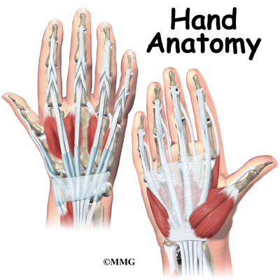
Hand Anatomy Dorsal
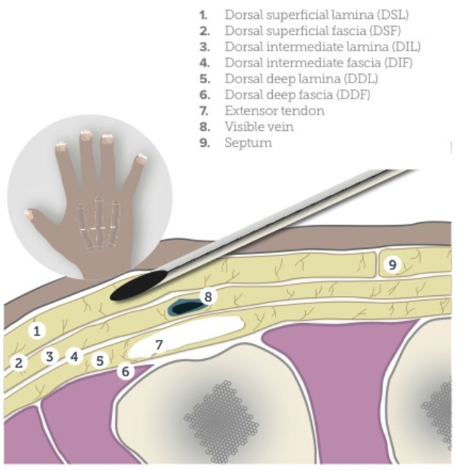
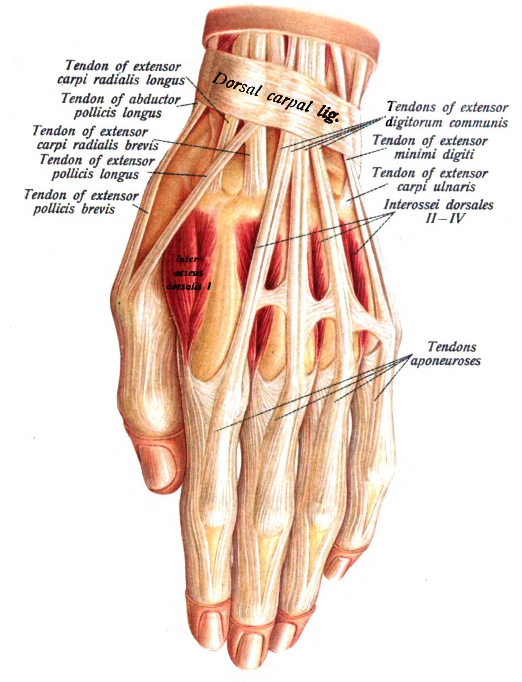
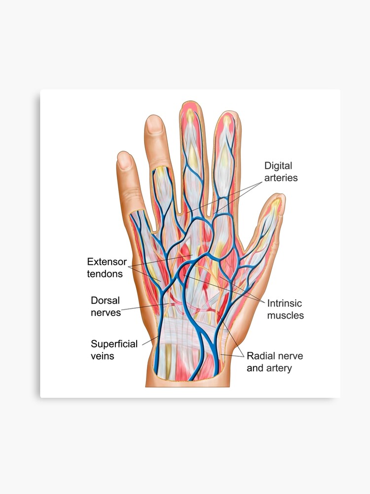
Most of what we can feel on the dorsal side of the hand is bone and most of what we can see are tendons along with a few superficial veins like the dorsal foot the dorsal hand has seven visible tendons although we dont always see them all at the same time and only one clearly visible muscle. Dorsal interossei the most superficial of all dorsal muscles these can be palpated on the dorsum of the hand.
 Hand Rejuvenation Aesthetics
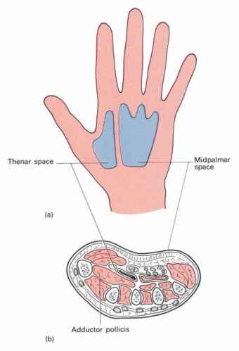
It is the area that sustains most pressure when using the palm of the hand for support such as in handstand.
Hand anatomy dorsal. Dorsal vs ventral dorsal is the backside while ventral is the opposite of backside. The connection point between the arm and the hand the wrist enables hand movements. For example the top of a dogs paw is its dorsal.
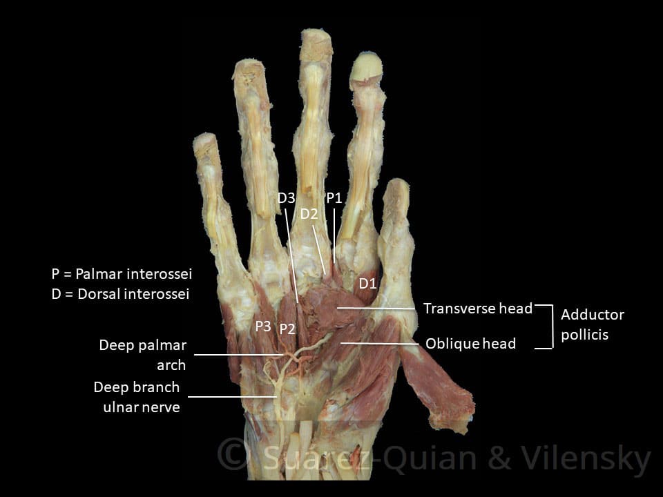
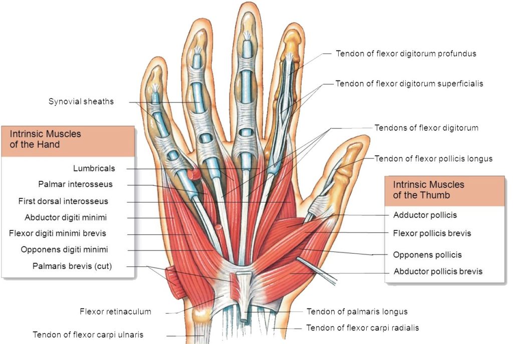
There are 4 dorsal and 3 palmar interossei muscles. Each interossei originates from the lateral and medial surfaces of the metacarpals. In invertebrates the nerve cord runs through the ventral side.
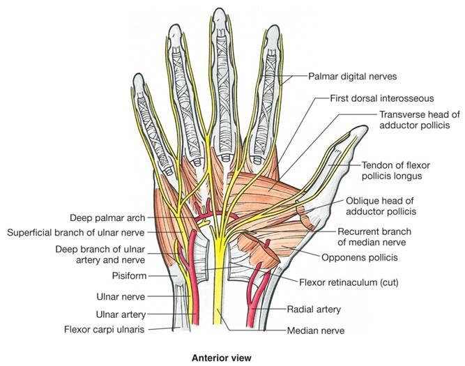
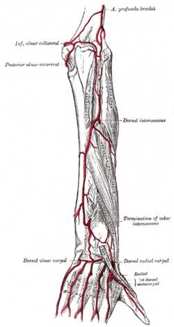
There are four dorsal interossei muscles. Palmar interossei adduct the fingers and dorsal interossei abduct the fingers hence paddab. The radial artery enters the hand by passing between the two heads of the first dorsal interosseous.
We think this is the most useful anatomy picture that you need. When a particular organ a is ventral to another b the organ b lies dorsal to the organ a. Hand dorsal view the human hand the most distal part of the upper limb is a remarkable feat of engineering and evolution.
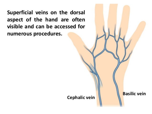
You can click the image to magnify if you cannot see clearly. The back of the hand shows the dorsal venous network a web of veins. The front or palm side of the hand is referred to as the palmar side.
The opisthenar area dorsal is the corresponding area on the posterior part of the hand. The heel of the hand is the area anteriorly to the bases of the metacarpal bones located in the proximal part of the palm. For improved clarity the directional term palmar from latin palma meaning palm of the hand is usually used to describe the front of the hand and dorsal is the back of the hand.
It is strong enough to allow climbers to tackle any mountain but also sufficiently precise for the manipulation of some of the worlds smallest objects and the performance of complex actions. The back of the hand is called the dorsal side. On the other hand the vertebrates have a ventral alimentary canal but a dorsal nerve cord.
This image added by admin. They insert onto the proximal phalanx and extensor hood of each finger.
 Spaces Of Hand Anatomy And Significance Bone And Spine
 Hand Anatomy Overview Bones Blood Supply Muscles Geeky

The Muscles Of The Hand Thenar Hypothenar Teachmeanatomy

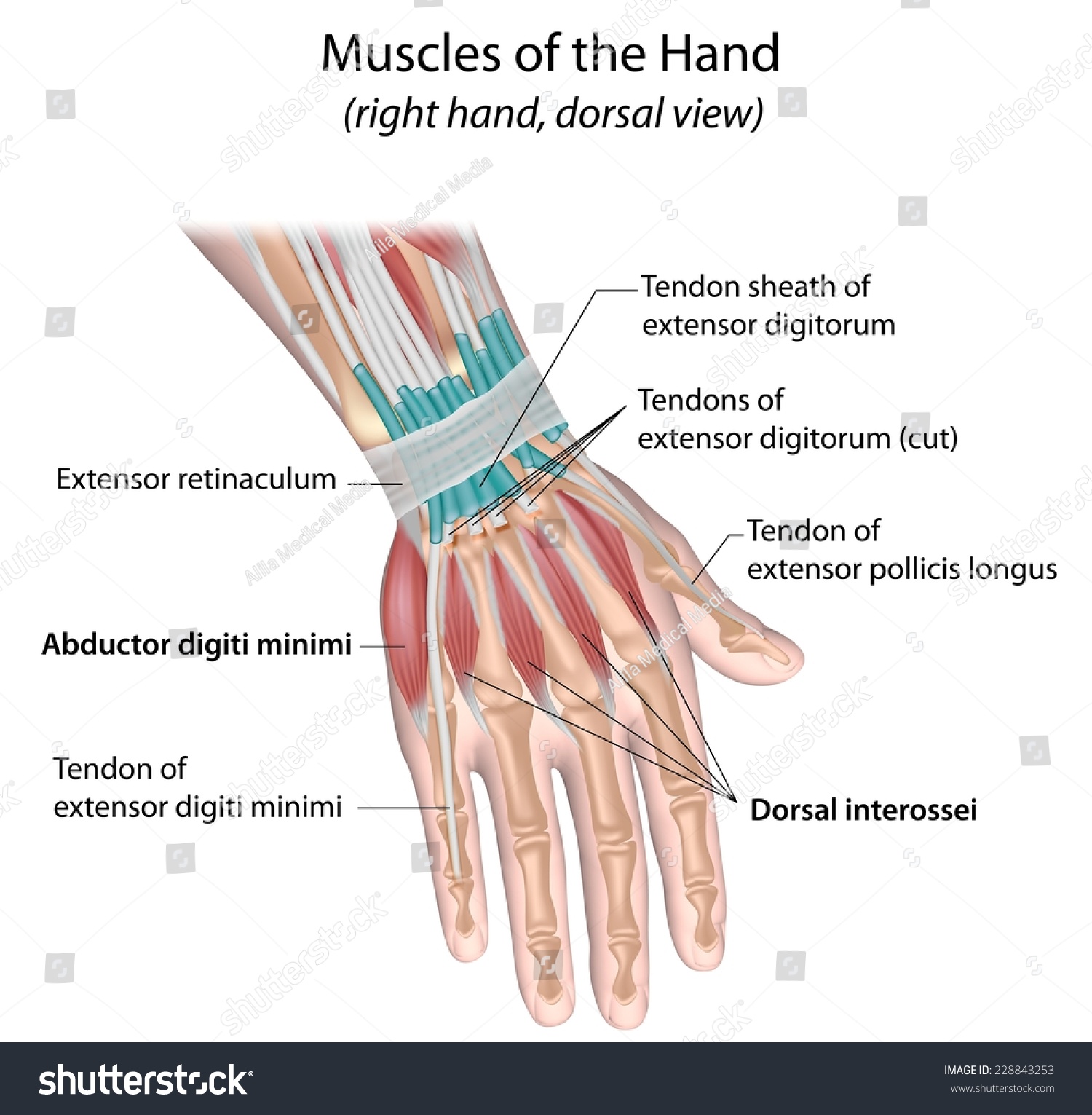 Muscles Hand Dorsal View Labeled Stock Illustration 228843253
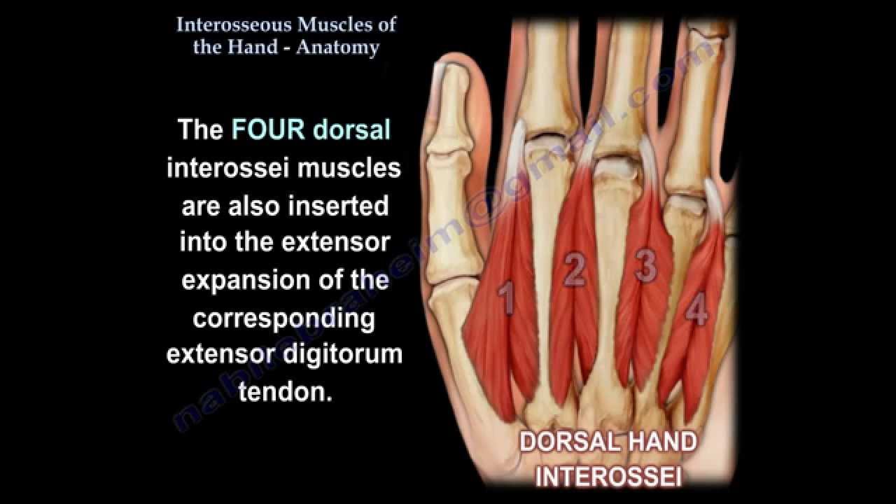 Interosseous Muscles Of The Hand Anatomy Everything You Need To Know Dr Nabil Ebraheim
 Wrist Hand Anatomy
 Muscles Of The Hand And Wrist Interactive Anatomy Guide
 File Hand Anatomy Jpg Wikimedia Commons
 Hand Anatomy And Function Bone And Spine
 Wrist Hand Anatomy
 Hand Anatomy Eorthopod Com
 Wrist Hand Atlas Of Anatomy
 Hand Muscles Attachment Nerve Supply Action Anatomy Info
 Structures Of The Hand Tendons Ligaments Teachmeanatomy

 Handcare Org Anatomy Muscles
 Anatomy Of Back Of Fore Arm And Dorsum Of Hand
 Superficial Radial Nerve Anatomy Orthobullets
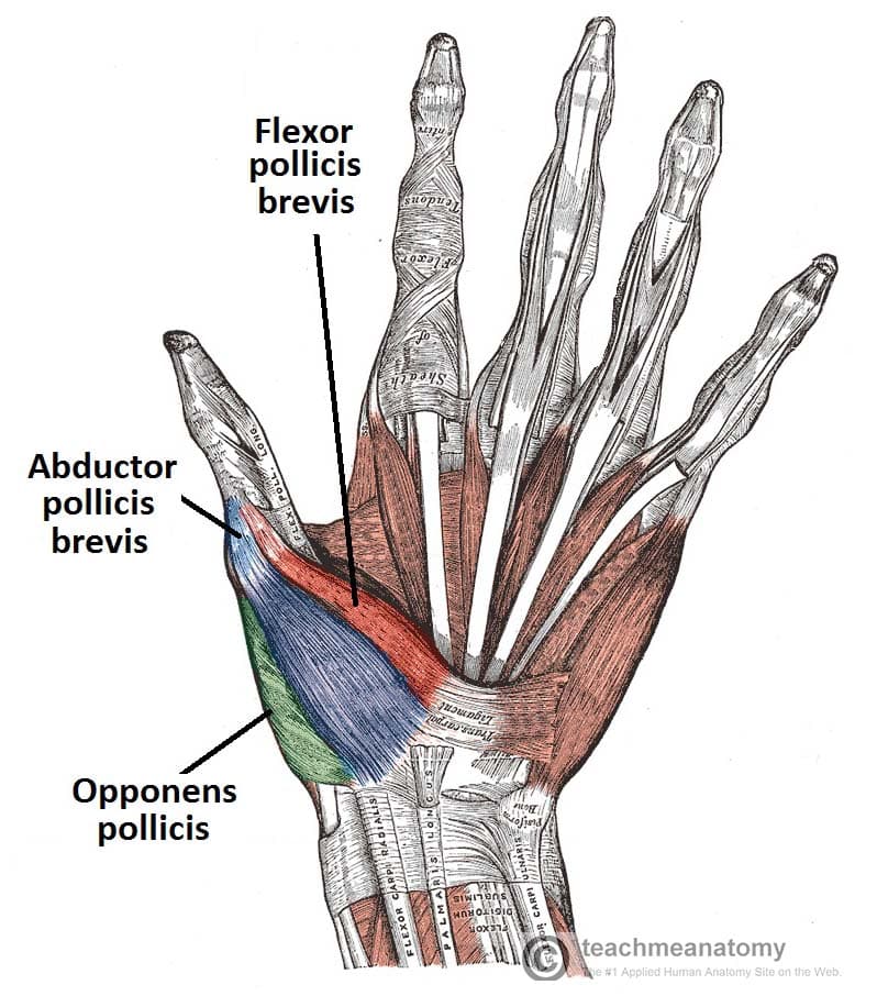 The Muscles Of The Hand Thenar Hypothenar Teachmeanatomy
 Understanding The Anatomy Of The Hand Health Life Media
 Dorsal Metacarpal Arteries Wikipedia
 Anatomy Of Back Of Human Hand Metal Print
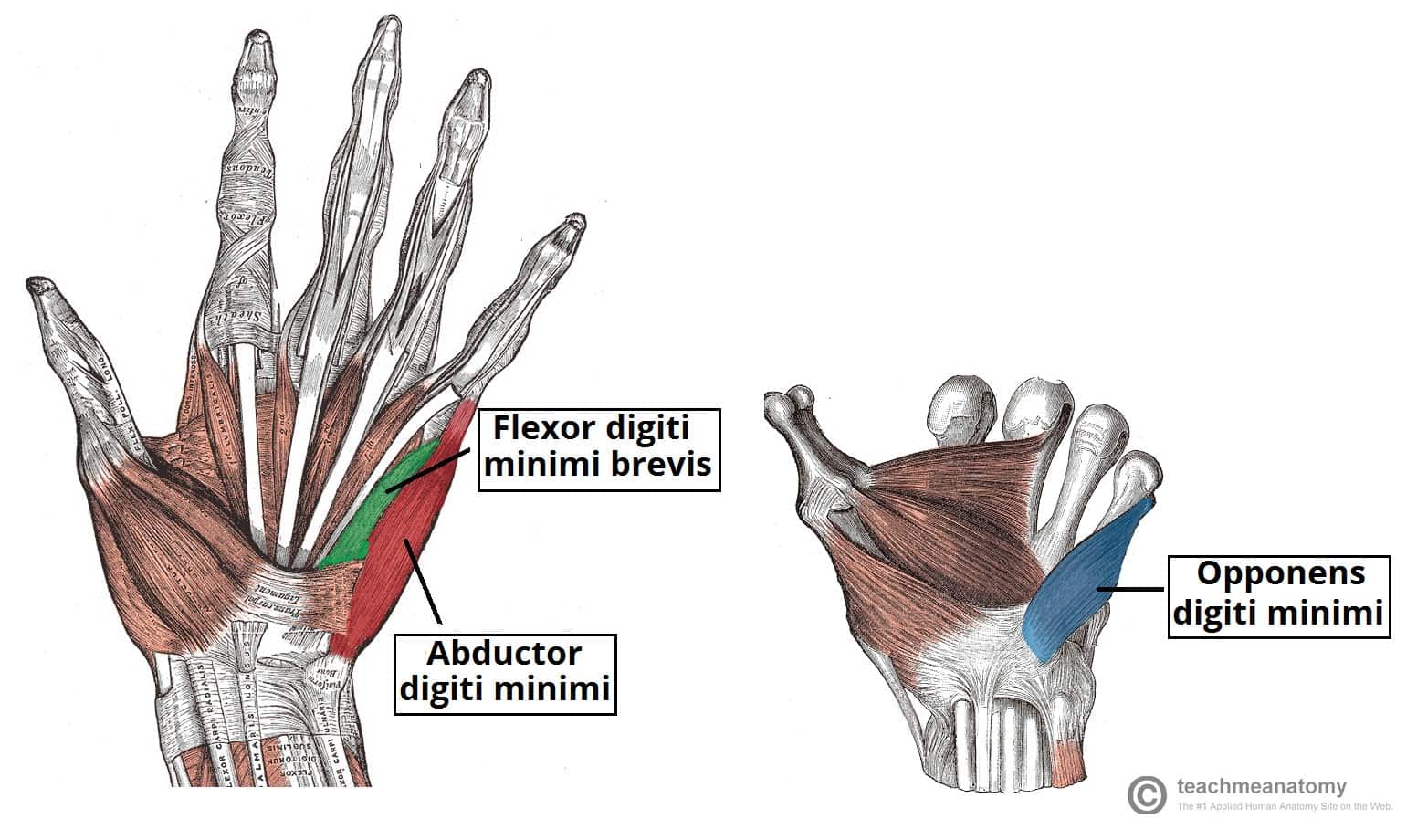 The Muscles Of The Hand Thenar Hypothenar Teachmeanatomy
Belum ada Komentar untuk "Hand Anatomy Dorsal"
Posting Komentar