Sural Anatomy
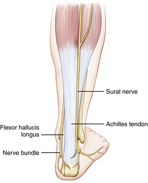
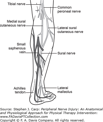
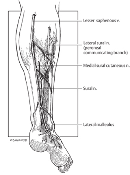
Sural nerve formation at the distal third of the gastrocnemius both sural cutaneous branches join to become the sural nerve descends on the posterolateral aspect of leg. The sural nerve is interposed between the one third part of the facial nerve segment and the periorbital branches that were tagged during the previous procedure.
 Entrapment Of The Lateral Cutaneous Nerve Of The Calf Bmj
Of or relating to the calf of the leg.
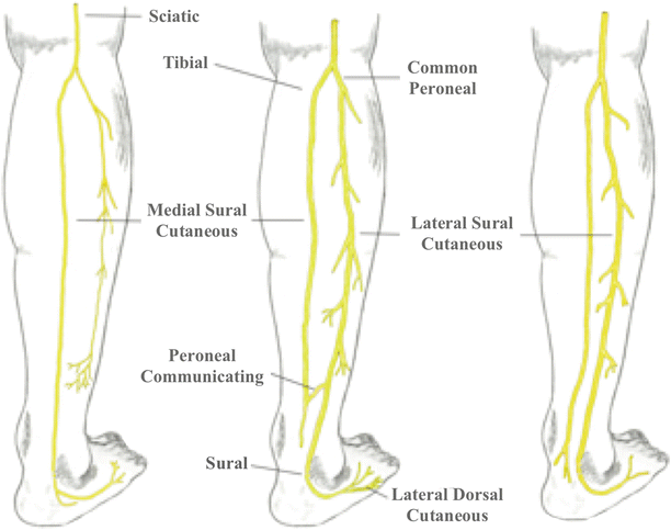
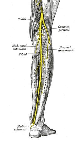
Sural anatomy. The sural nerve is a guide to the parent trunk which is the tibial nerve. The term applies to any of four or five arteries arising from the popliteal artery with distribution to the muscles and integument of the calf and with anastomoses to the posterior tibial medial and lateral inferior genicular arteries. The sural nerve is a sensory nerve of the lower limb formed by the union of branches from the tibial nerve as well as common fibular nerve supplying sensation to the lower lateral aspect of the calf and foot.
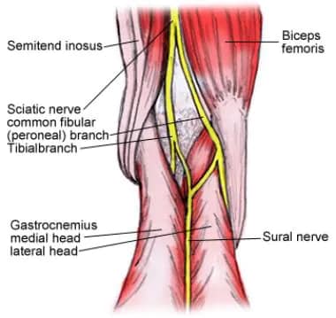
It is made up of branches of the tibial nerve and common fibular nerve the medial cutaneous branch from the tibial nerve and the lateral cutaneous branch from the common fibular nerve. The short saphenous nerve initially courses posterior between the heads of the gastrocnemius muscle. It travels within subcutaneous tissue adjacent to the small saphenous vein in the lower posterolateral calf.
The tibial nerve can be located by tracking the sural nerve proximally. Content related to sural 8 ways to say yes yes means yes but so do all of these words. This is in concordance with its course as it passes in the lateral side of the lower calf ankle and foot.
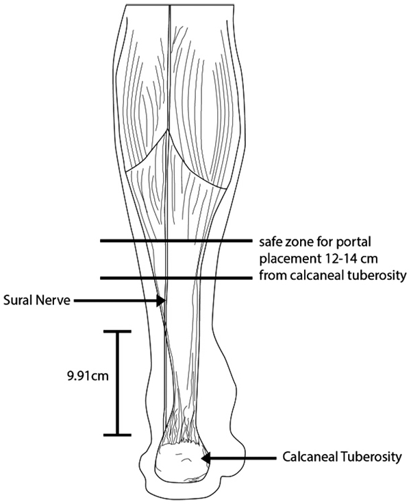
High in the popliteal fossa the sciatic nerve divides into its two main branches on route to serve the leg namely the tibial nerve and the common fibular nerve. Intraoperative problems may include damage to the sural nerve which is usually between 7 and 14 mm posterior to the tip of the fibula. It also transmits sensations from the 5 th toe.
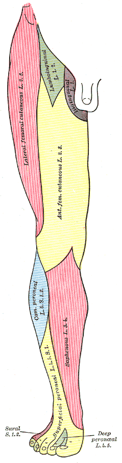
The sural nerve is responsible for the sensation of the skin of the lateral foot and lateral lower ankle. The sural nerve is a sensory nerve made up of collateral branches off of the common tibial and common fibular nerve. Sural nerve anatomy as aforesaid it is purely a sensory nerve and does not consist of motor fibers.
It is a purely sensory nerve. Sural means related to the calf. The sural nerve is a sensory nerve in the calf region of the leg.
The sural nerve passes down to the posterolateral.

 Popliteal Nerve Block Background Indications
 Pediagenosis
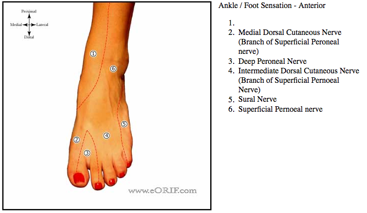 Ankle Anatomy Eorif
 Sural Nerve Entrapment Springerlink
 Lateral Sural Cutaneous Nerve Wikidoc
 Anatomy Evaluation And Operative Setup For Posterior Ankle
 Tibial Nerve Neurologyneeds Com
 Cutaneous Nerve Blocks Of The Lower Extremity Nysora
 The Sural Communicating Branch Stock Illustration
 Cadaveric Anatomical Study Of Sural Nerve Where Is The Safe
 Tibial Nerve Seriously Sciatic
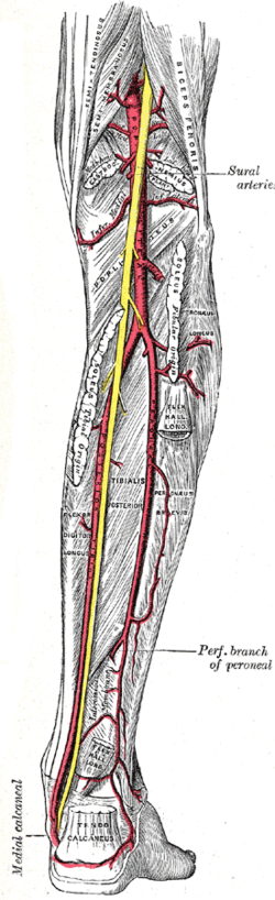 Sural Arteries Wikipedia
 Microsurgery Medial Sural Artery Perforator Msap Flap Harvest
 What Causes Sural Nerve Compression And How To Manage It
Sural Nerve Entrapment Springerlink

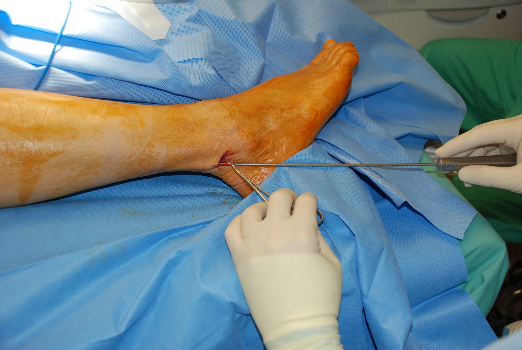 Sural Nerve Graft Harvest Iowa Head And Neck Protocols
 Arterial System Of The Leg
 Pdf Sural Nerve Lipoma An Anatomy Review Semantic Scholar
 Sural Nerve Knee Preservation Foundation
 Entrapment Neuropathies In The Foot And Ankle Peripheral
 The Medial Sural Artery Perforator Island Flap As A Simpler
![]() Common Peroneal Nerve Physiopedia
Common Peroneal Nerve Physiopedia
 Sural Nerve Injury And Neuroma Neupsy Key

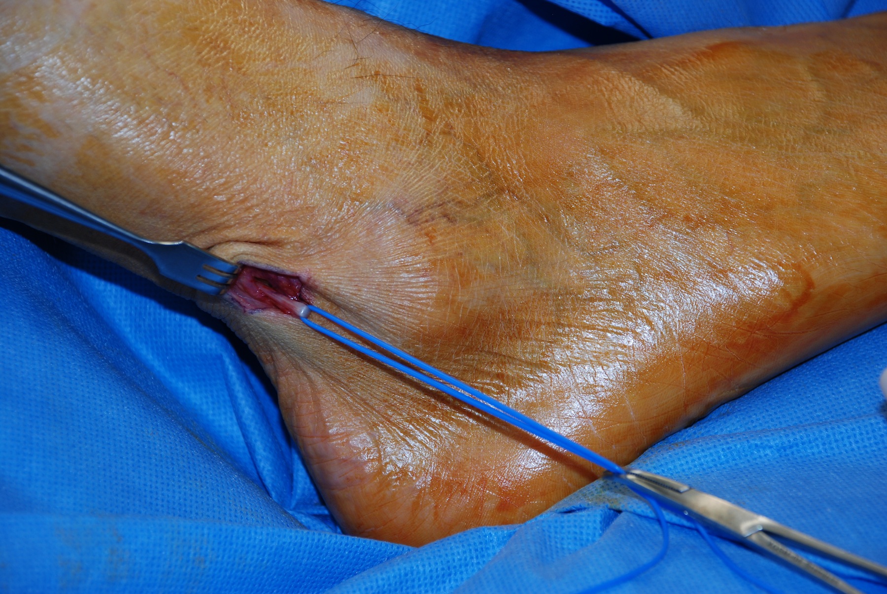 Sural Nerve Graft Harvest Iowa Head And Neck Protocols
Belum ada Komentar untuk "Sural Anatomy"
Posting Komentar