Heart Anatomy Ct
It is very important in determining the relationship of the coronary artery to the aorta and the pulmonary artery. Click on different parts of the heart and coronary vessels on this axial ct and answer corresponding questions.
 Left Dominant Coronary Circulation Radiology Case
It can also detect aneurysm of the pulmonary artery below and of the aorta.

Heart anatomy ct. A heart ct scan is used to view your heart and blood vessels. Cardiac ct is a heart imaging test that uses ct technology with or without intravenous iv contrast dye to visualize the heart anatomy coronary circulation and great vessels which includes the aorta pulmonary veins and arteries. With these scanners the heart and coronary arteries are routinely imaged as a motion free volume of data.
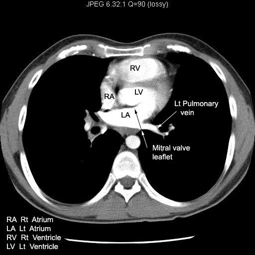
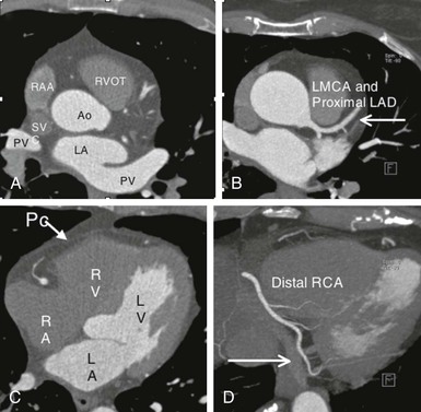
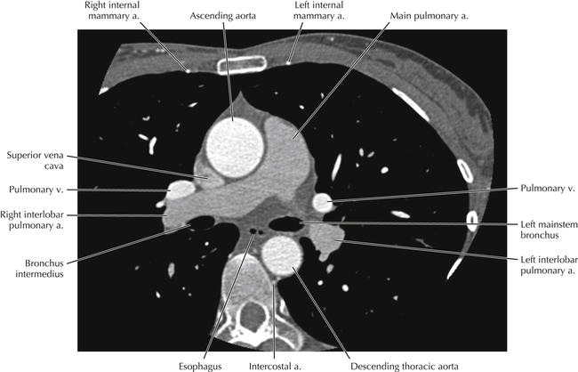
The advent of multidetector computed tomography ct particularly with scanners having 64 or more detectors has continued to improve temporal resolution and allows the acquisition of isotropic voxels. Typically cardiac ct axial examinations are specifically tailored to image the heart and therefore the imaging volume is restricted to only a portion of the thorax. Often the examination begins just below the carina as this first plate shows.
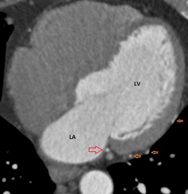
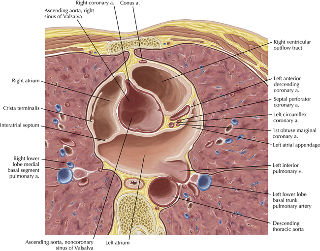
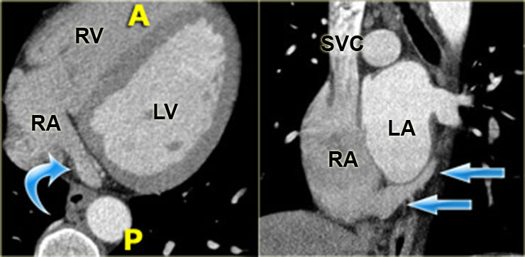
The crista terminalis is a vertical fibromuscular ridge that separates the smooth portion of the right atrium which receives the superior and inferior vena cavae and coronary sinus from the right atrial appendage and the remainder of the right atrium containing pectinate muscles. Anatomy of the heart coronary ct interactive atlas of the human body using cross sectional imaging in this interactive anatomy atlas of the human heart the anatomical structures are visible on a contrast materialenhanced computed tomography ct of the heart and coronary arteries. Cardiac ct is one of the most useful technique in evaluating the origin and course of anomalous coronary arteries.
It can diagnose birth defects buildup of plaque that may be blocking arteries and tumors. Anatomy of the heart quiz ct click on the image description. Computed tomography of the heart or cardiac ct is routinely performed to gain knowledge about cardiac or coronary anatomy to detect or diagnose coronary artery disease cad to evaluate patency of coronary artery bypass grafts or implanted coronary stents or to evaluate volumetry and cardiac function including ejection fraction.
![]() Left Transaxial Ct Images Reveal The Cardiac Anatomy
Left Transaxial Ct Images Reveal The Cardiac Anatomy
 The Radiology Assistant Cardiac Anatomy
![]() Left Transaxial Ct Images Reveal The Cardiac Anatomy
Left Transaxial Ct Images Reveal The Cardiac Anatomy
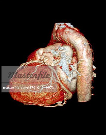 Normal Heart 3d Ct Scan Stock Photo Masterfile
 Basic Thoracic Anatomy And Physiology The Core Curriculum
 Ct Scan Show Cardio Heart Anatomy Stock Image Download Now
 Superior Specificity In Cardiac Ct Siemens Healthineers India
The Radiology Assistant Cardiac Anatomy
 Cross Sectional Anatomy Of The Body Mdct Atlas Ctisus Com
 Cardiac Anatomy Using Ct Radiology Key
 Cardiac Computed Tomography Thoracic Key

 Anatomy Of The Heart And Coronary Arteries Coronary Ct
 Advances In Cardiac Ct Technology Daic
 The Radiology Assistant Cardiac Anatomy
 The Radiology Assistant Cardiac Anatomy
 Chest Ct Anatomy Radiology Key
 The Radiology Assistant Cardiac Anatomy
 Cardiac Anatomy Using Ct Radiology Key
 Figure3 A Contrast Ct Of The Frontal Plane Showing The
 Thorax Of The Dog Cross Sectional Anatomy On Computed
 Cross Sectional Cardiac Anatomy
 Science Source Internal Heart Anatomy 3d Ct Scan

Belum ada Komentar untuk "Heart Anatomy Ct"
Posting Komentar