Anatomy Pregnancy
The goal of maternity care is a healthy pregnancy with a physically safe and emotionally satisfying outcome for mother infant and family. These are all normal and natural questions and in fact knowing what your fetus looks like and understanding how she is developing may even help you feel closer to your growing baby.
 Anatomy Of A 40 Week Pregnant Woman Ligaments Not Shown
The anatomy scan is a level 2 ultrasound which is typically performed on pregnant women between 18 and 22 weeks.

Anatomy pregnancy. There is no real separation between the areas of your pelvis and abdomen. Anatomy to better understand the changes your body goes through during the last trimester and labor it is helpful to be familiar with basic anatomy. By the 20th week of pregnancy the baby can weigh up to 11 ounces and measure 10 inches outstretched.
When the pregnancy hits the 20th week of gestation an anatomy ultrasound is often ordered. Consistent health supervision and surveillance are of utmost importance. One greys anatomy doctor is pregnant after the season 16 premiere.
Those who want to can find out the sex of the baby if desired. Heres why you should have see the surprise coming and its full backstory. As a pregnant woman or the partner of one you may be curious about the anatomy of a pregnancy the fetus and the special organs that keep it happy and healthy and connected to mom.
If you have a condition that needs to be monitored such as carrying multiples you may have more than one detailed ultrasound. This sonogram is used to determine fetal anomalies the babys size and weight and also to measure growth to ensure that the fetus is developing properly. Most anatomy scans are performed in the second trimester of pregnancy typically at 20 weeks but they can be done anytime between 18 weeks and 22 weeks.
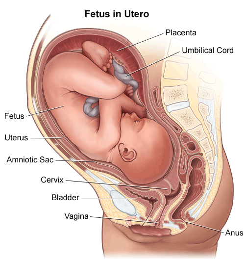
The beautiful and efficient anatomy of pregnancy. Conception to birth visualized by alexander tsiaras. In the picture here you can see that the vagina is behind the bladder sac that collects urine and urethra tube for moving urine out of bladder and body.
Medical anatomical pregnant human female pelvis with pregnancy 9 months baby fetus model life size with removable organs 4 parts hand painted 45 out of 5 stars 2 11255 112. Open the activity on the right to compare your body before pregnancy to your body at 37 weeks. Before pregnancy most of the space in your abdomen is taken up by the large and small intestines.
Youll see how your body adjusts in amazing ways to support your growing baby. When a level 2 ultrasound is done. However many maternal adaptations are unfamiliar to pregnant women and their families.
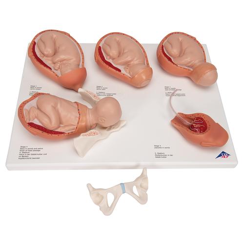 Labor Stages Model Small 3b Smart Anatomy
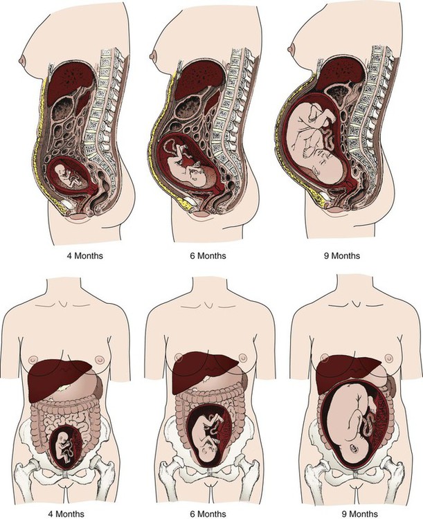 Anatomy And Physiology Of Pregnancy Nurse Key
 Uterus Growth During Pregnancy
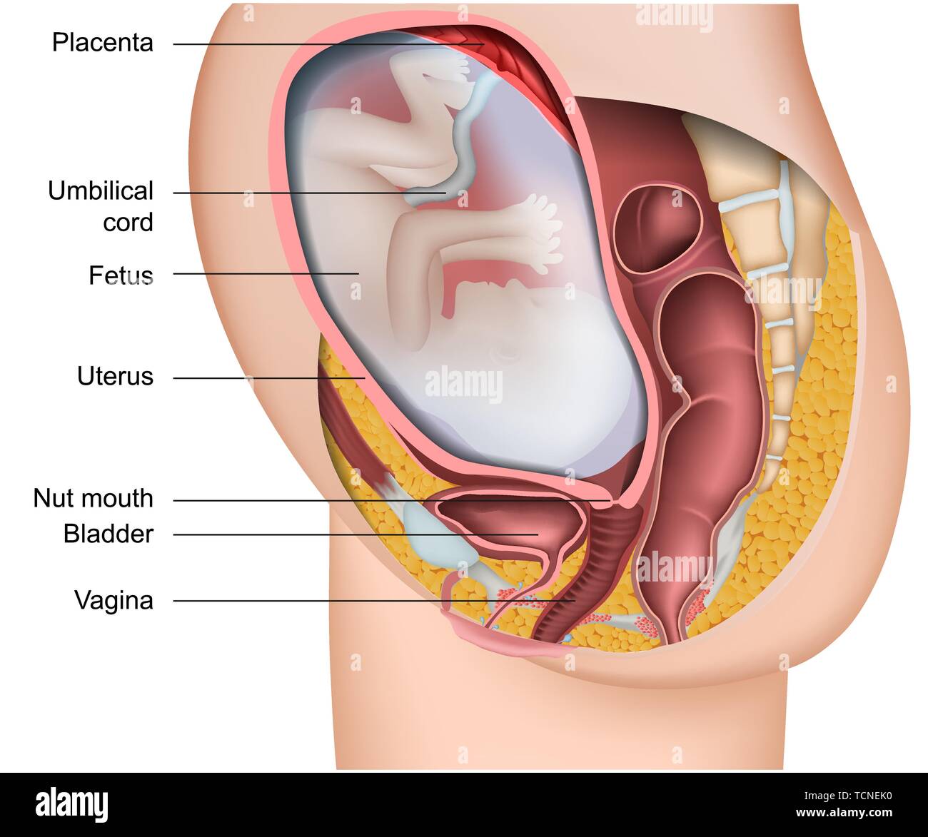 Pregnancy 3d Medical Vector Anatomy Illustration Isolated On
 Science Source Pregnancy Anatomy 15th Century Artwork

 Pregnancy Anatomy Illustration On Behance
 Clip Art Vector Normal Pregnant Female Anatomy Stock Eps
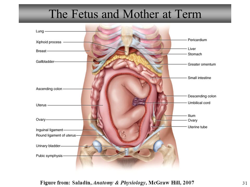 Anatomy And Physiology Pregnancy Growth And Development
 Anatomy Of Pregnancy Medivisuals Medical Illustration
 4d Human Pregnancy Pelvis Anatomy Body Anatomical Teaching Model 4893409260603 Ebay

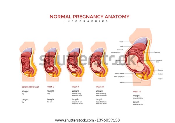 Normal Pregnancy Anatomy Medical Infographic Chart Stock
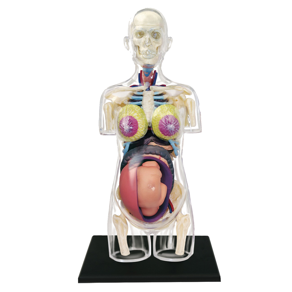 4d Transparent Pregnancy Model
 Pregnancy Motion Anatomy By Usamau04 Melambe
 Amazon Com Ahawoso Outdoor Garden Flag 12x18 Inches

 How Does Your Anatomy Change During Pregnancy Socratic
 Pregnant Woman Colorful Anatomy Isolated Stock
 Breast Cancer Treatment During Pregnancy
 This Horrifying Animation Shows You How A Woman S Organs Move During Pregnancy




Belum ada Komentar untuk "Anatomy Pregnancy"
Posting Komentar