Pelvic Bone Anatomy
There are four articulations within the pelvis. The pelvic region of the trunk is the lower part of the trunk between the abdomen and the thighs.
 Skeleton Pixel Art Skull And Bones Stock Vector
The bones of the pelvis and lower back work together to support the bodys weight anchor the abdominal and hip muscles and protect the delicate vital organs of the vertebral and abdominopelvic cavities.

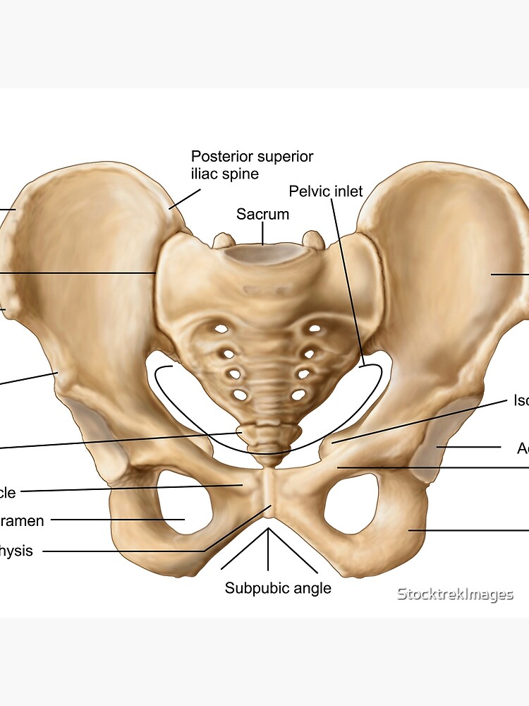
Pelvic bone anatomy. It includes the following structures. Sacrococcygeal symphysis between the sacrum and the coccyx. Anatomy of the pelvis includes anatomy of the bony pelvis and its contents.
Pubic symphysis between the pubis bodies of the two hip bones. The hip bones join to the upper part of. The gap enclosed by the bony pelvis called the pelvic cavity is the section of the body underneath the abdomen and mainly consists of the reproductive organs sex organs and the rectum while the pelvic floor at the base of the cavity assists in supporting the organs of the abdomen.
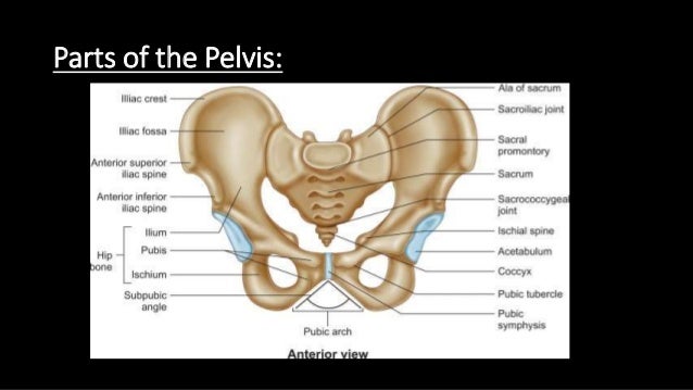
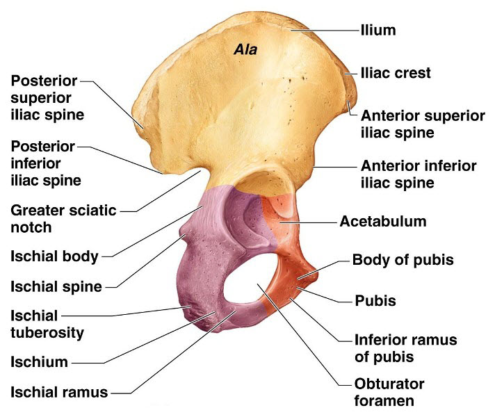
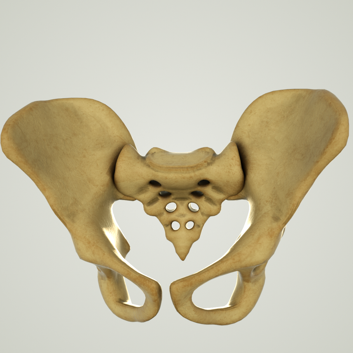
Bony pelvis or pelvic skeleton is formed by hip bones sacrum and coccyx. There are three bones of the pelvis. The pelvis is formed by four bones which include a pair of hip bones otherwise known as innominate bones.
The external male genitals include the penis scrotum and testicles. These bones connect the axial skeleton to the lower limbs and therefore play a role in bearing the weight of the upper body. These foramina are created by the positioning of bony.
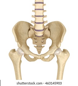
Explore and learn about the pelvis with our 3d interactive anatomy atlas. These bones also act as attachments for many muscles and ligaments within the pelvis and lower limbs. Anatomy pelvis also called bony pelvis or pelvic girdle in human anatomy basin shaped complex of bones that connects the trunk and the legs supports and balances the trunk and contains and supports the intestines the urinary bladder and the internal sex organs.
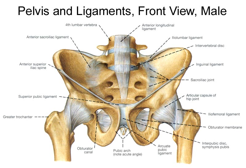
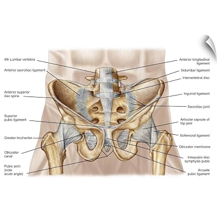
Together they form the part of the pelvis called the pelvic girdle. The two main ligaments of the pelvis are the sacrotuberous and sacrospinous ligaments. Sacroiliac joints x2 between the ilium of the hip bones and the sacrum.
The hip bone sacrum and coccyx. There are two hip bones one on the left side of the body and the other on the right. The hip joint is a ball and socket joint created by the femur and a part of the pelvis called the acetabulum.
In this article we would discuss bony pelvis. Bones of the pelvis and lower back. The vertebral column of the lower back includes the five lumbar vertebrae the sacrum and the coccyx.
This joint and its ability to rotate in many angles is one of many pieces of anatomy that allows humans to walk.
 Skeleton Anatomy Human Skeletal System Cross Section Bones
 Pelvis Human Skeleton Female Pelvis Bone Anatomy Hip
 Pelvis Wikipedia
 Bony Pelvis Anatomy Bone And Spine
 Anatomy Of Human Pelvic Bone Duvet Cover
 Hip Bones Anatomy Os Coxae Pelvic Girdle Ilium Ischium
 Pelvic Bones Anatomy Male Vs Female Pelvis
 Amazon Com Canvas On Demand Anatomy Of Human Pelvic Bone
 Anatomy Of Human Pelvic Bone And Ligaments Print Wall Art
The Pelvis Anatomy Images Pelvic Floor Connective Tissues
 Pelvic Bone Stock Photos And Images 123rf
 Hip Bone Anatomy Or Pelvic Bone Ilium Pubis Ischium Bone
 Human Pelvic Bone Spine Structure
 Pelvis Hip Anatomy
 Pelvis Anatomy Recon Orthobullets
 Pelvic Bone
 Anatomy Of Human Pelvic Bone Acrylic Print
 Pelvis Bones And The Ligaments Front On And Rear View
 8 3 The Pelvic Girdle And Pelvis Anatomy And Physiology
 The Pelvis Human Anatomy And Physiology Lab Bsb 141
 Vector Illustration Female Pelvis Bone Anatomy Eps
 Musculoskeletal Pelvic Anatomy Sciencedirect
 Pelvic Bone Images Stock Photos Vectors Shutterstock
 Pelvic Bone Anatomy Revision From 1st Year Diagram Quizlet
 The Hip Bone Human Anatomy
 Anatomy Of Human Pelvic Bone Tote Bag
 Vector Illustration Female Pelvis Bone Anatomy Eps
 Anatomy Human Pelvic Bone Wall Mural Wallmonkeys Com
Belum ada Komentar untuk "Pelvic Bone Anatomy"
Posting Komentar