Pelvic Anatomy Bones
The hip bone or coxal bone forms the pelvic girdle portion of the pelvis. Explore and learn about the pelvis with our 3d interactive anatomy atlas.
The pelvic cavity is a body cavity that is bounded by the bones of the pelvis and which primarily contains reproductive organs and the rectum.

Pelvic anatomy bones. Evolutionary scientists believe this stems from mans hunter roots as a leaner pelvis made running easier. The human pelvis is a group of bones fused together and functions to join the axial skeleton bones of the head neck and vertebrae to the lower appendicular skeleton. A distinction is made between the lesser or true pelvis inferior to the terminal line and the greater or false pelvis above it.
Bones of the pelvis and lower back. Anatomy pelvis also called bony pelvis or pelvic girdle in human anatomy basin shaped complex of bones that connects the trunk and the legs supports and balances the trunk and contains and supports the intestines the urinary bladder and the internal sex organs. The pelvic girdle is formed by a single hip bone.
The pelvic cavity is a body cavity that is bounded by the bones of the pelvis and which primarily contains reproductive organs and the rectum. A distinction is made between the lesser or true pelvis inferior to the terminal line and the greater or false pelvis above it. The pelvis is formed by four bones which include a pair of hip bones otherwise known as innominate bones.
The bones of the pelvis and lower back work together to support the bodys weight anchor the abdominal and hip muscles and protect the delicate vital organs of the vertebral and abdominopelvic cavities. Together they form the part of the pelvis called the pelvic girdle. The bones of the pelvis are the hip bones.
The hip bone attaches the lower limb to the axial skeleton through its articulation with the sacrum. The pelvic bones are smaller and narrower. There are two hip bones one on the left side of the body and the other on the right.
The vertebral column of the lower back includes the five lumbar vertebrae the sacrum and the coccyx. The right and left hip bones plus the sacrum and the coccyx together form the pelvis. These foramina are created by the positioning of bony.
The two main ligaments of the pelvis are the sacrotuberous and sacrospinous ligaments. The axial skeleton joining the pelvis is the spinal column specifically the lumbar spine and the bones of the coccyx.
 Anatomy Of The Horse Osteology
 Bones Of Pelvis Anatomy Lecture Slides Docsity
 Pelvis Definition Anatomy Diagram Facts Britannica
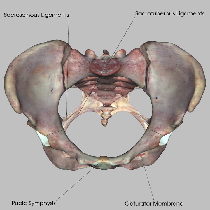 Bones And Ligaments Of The Pelvis
 Bones Of The Pelvis And Lower Back
 Anatomy Of Human Pelvic Bone By Stocktrek Images Canvas Print
 Pelvis Anatomy Bone Abdomen Human Body Sacrum
 Bones Of The Pelvis Anatomy Hip Bone Anatomy 1
 Human Male Anatomy Scheme Main Pelvic Bones Vector Illustration
 The Pelvic Girdle Of Human Hip Bone Anatomy Vector

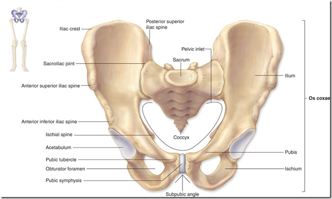 The Bones Of The Pelvis And Lower Back Anatomy Medicine Com
 Anatomy Of Human Pelvic Bone By Stocktrek Images Canvas Print
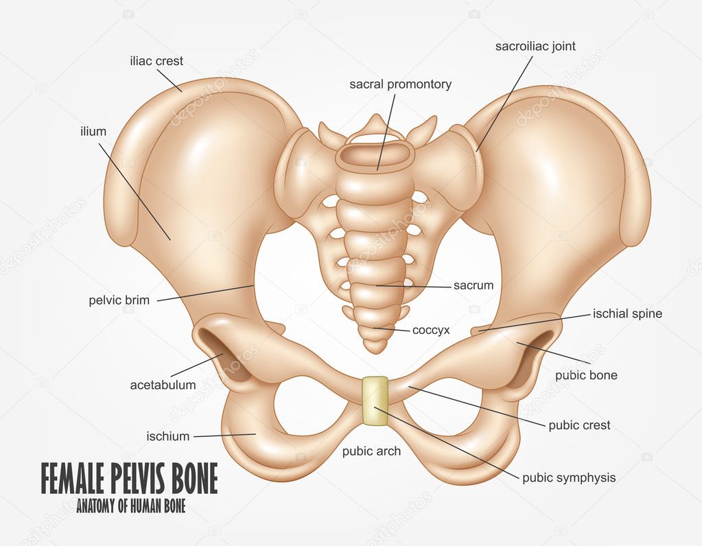 Diagram Of Female Pelvic Bone Female Pelvis Bone Anatomy
:watermark(/images/watermark_5000_10percent.png,0,0,0):watermark(/images/logo_url.png,-10,-10,0):format(jpeg)/images/atlas_overview_image/987/xSiX0lUlLxEIjBI8malVw_bones-pelvis-femur_english.jpg) Pelvis Anatomy Bones Joints Ligaments And Foramina Kenhub
Pelvis Anatomy Bones Joints Ligaments And Foramina Kenhub
 Hip Bones Anatomy Os Coxae Pelvic Girdle Ilium Ischium
Pelvic Girdle Human Anatomy Organs
 Anatomy Of Human Pelvic Bone Canvas Print
 Pelvic Girdle Hip Bone Muscle Anatomy Human Anatomy
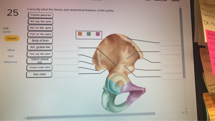 Solved Correctly Label The Bones And Anatomical Features
:background_color(FFFFFF):format(jpeg)/images/library/11036/muscles-pelvis-hip-femur_english.jpg) Hip And Thigh Bones Joints Muscles Kenhub
Hip And Thigh Bones Joints Muscles Kenhub
 Hip Bones Anatomy Os Coxae Pelvic Girdle Ilium Ischium
Pelvic Anatomy And Function Bone Rhythms Human Kinetics
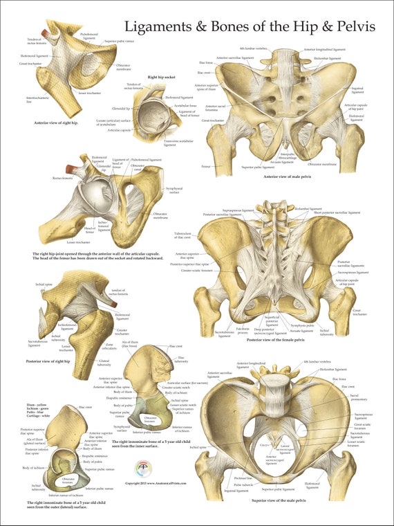 Human Ligaments And Bones Of The Hip And Pelvis Anatomy Poster 18 X 24 Medical Chart
 Pelvic Bone Anatomy Anatomy Of Pelvic Girdle In This Image
 Home Pelvis Anatomy Anatomy Bones Hip Anatomy
 The Pelvis Anatomy Images Pelvic Floor Connective Tissues
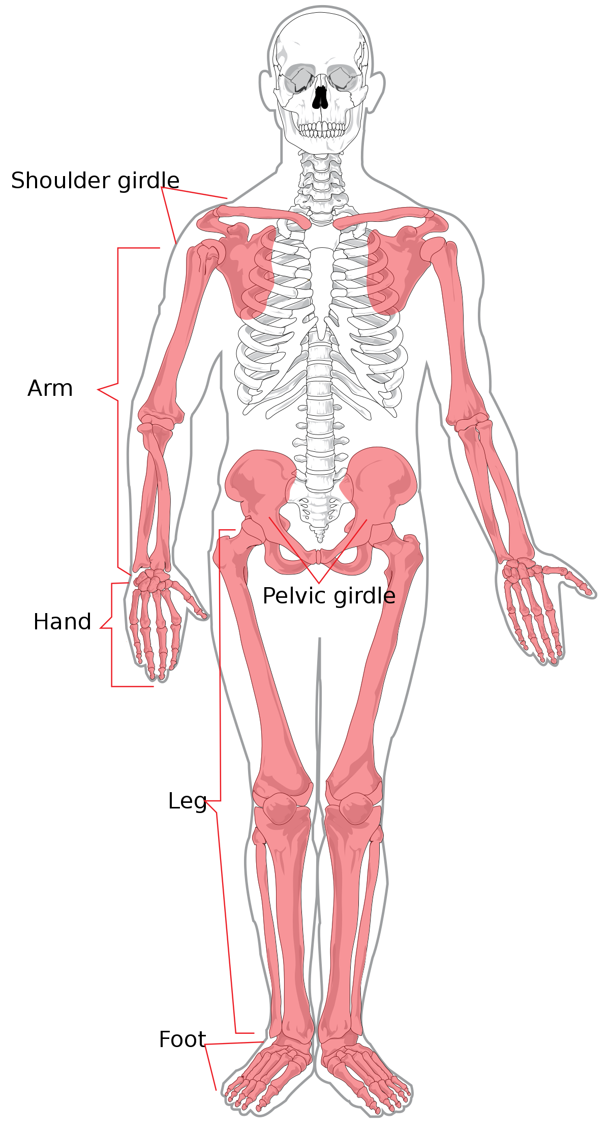 Appendicular Skeleton Wikipedia



Belum ada Komentar untuk "Pelvic Anatomy Bones"
Posting Komentar