Heart Anatomy Diagram
Start studying heart anatomy. This amazing muscle produces electrical impulses that cause the heart to contract.
Lots of mito the pericardium is a double walled sac containing the heart an describe the location of the heart in t located in the center of the chest behind the sternum.

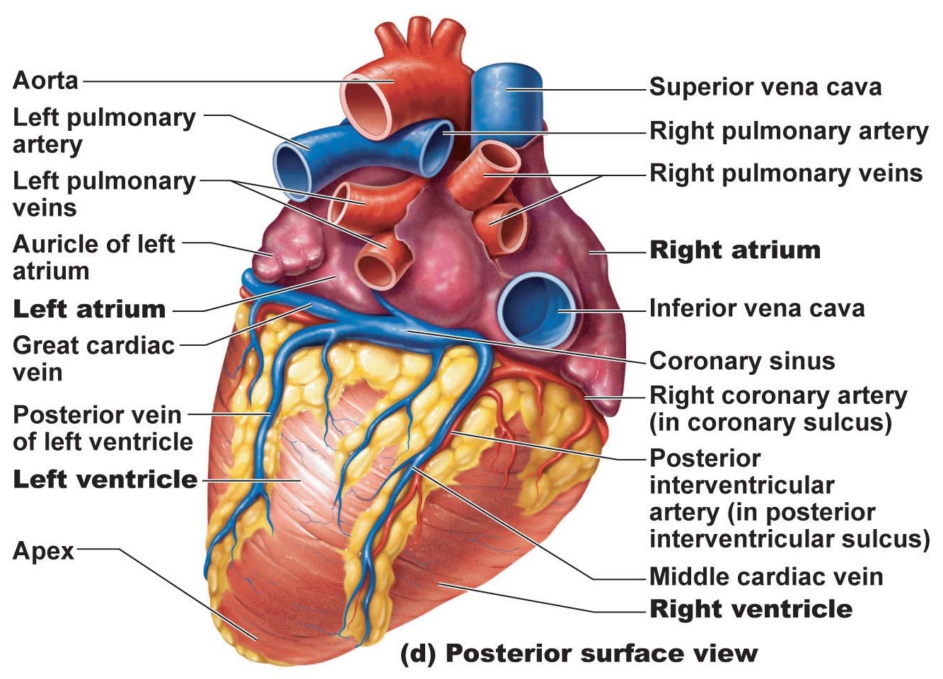
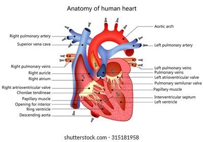
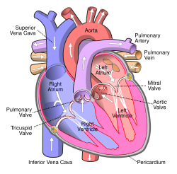
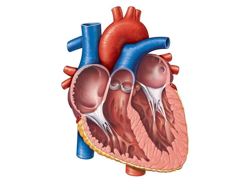
Heart anatomy diagram. The heart is a mostly hollow muscular organ composed of cardiac muscles and connective tissue that acts as a pump to distribute blood throughout the bodys tissues. The heart pumps blood through the network of arteries and veins called the cardiovascular system. Structure of heart wall in human heart diagram epicardium.
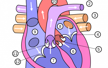
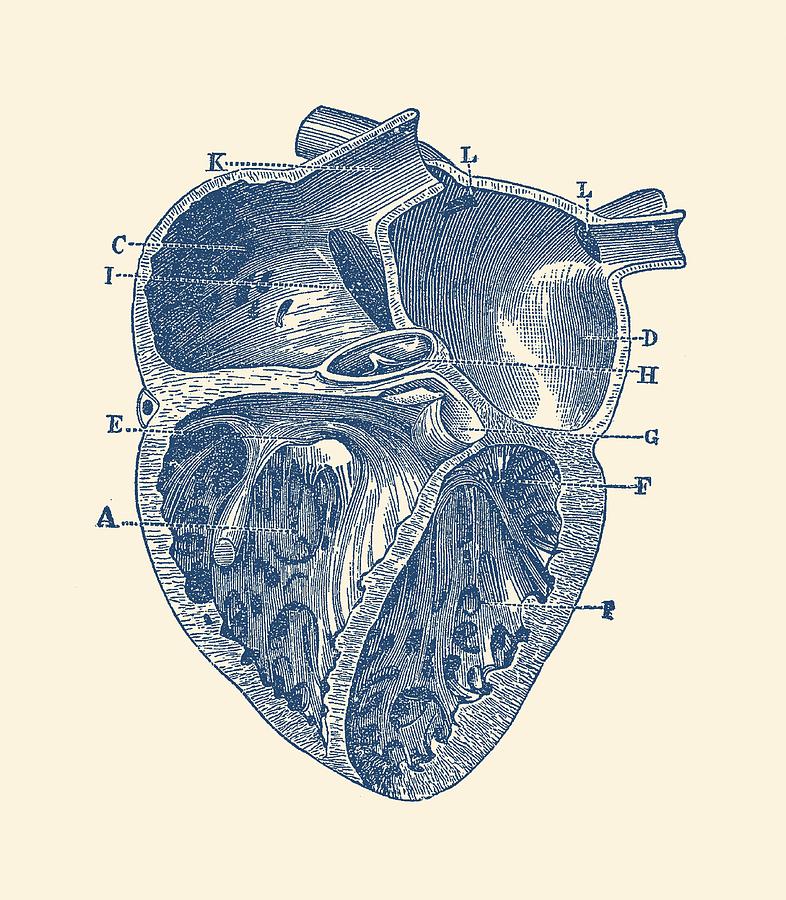
The heart has four chambers. Located in the center of the chest behind the sternum or the short fibers branchedstriated. Using our unlabeled heart diagrams you can challenge yourself to identify the individual parts of the heart as indicated by the arrows and fill in the blank spaces.
The right atrium receives blood from the veins and pumps it to the right ventricle. The heart is situated within the chest cavity and surrounded by a fluid filled sac called the pericardium. The outer layer of the pericardium surrounds the roots of your hearts major blood vessels and is attached by ligaments to your spinal column diaphragm and other parts of your body.
The heart sits within a fluid filled cavity called the pericardial cavity. The heart an image of the heart with blank labels attached the circulatory system upper body image with blank labels attached the circulatory system lower body image with blank labels attached the circulatory system a pdf file of the upper and lower body for printing out to use off line. The walls and lining of the pericardial cavity are a special membrane known as the pericardium.
The anatomy of the heart. The right ventricle receives blood from the right atrium and pumps it to the lungs where it is loaded with oxygen. Anatomy of the heart pericardium.
This exercise will help you to identify your weak spots so youll know which heart structures you need to spend more time studying with our heart quizzes. It is the simple squamous endothelium layer which lines the inside of the. A double layered membrane called the pericardium surrounds your heart like a sac.
The heart is the epicenter of the circulatory system which supplies the body with oxygen and other important nutrients needed to sustain life. It is divided by a partition or septum into two halves and the halves are in turn divided into four chambers. Because the heart points to the left about 23 of the hearts mass is found on the left side of the body and the other 13 is on the right.
Learn vocabulary terms and more with flashcards games and other study tools. The myocardium is muscular middle layer of heart wall which contain. The epicardium is one of the most outer layers of the heart wall.
1 or 2 nuclei.
 Heart Anatomy Anatomy And Physiology
 Human Anatomy Heart Color Vintage
 Human Heart Anatomy Free Online Game Biology Helpful Games
 Heart Anatomy Chambers Valves And Vessels Anatomy
Cardiovascular System Of The Dog
 Inner Heart Diagram Vintage Anatomy
 Pandarllin Throw Pillow Cover Scientific Anatomical Diagram Human Heart Anatomy Life Science Body Cardiac Muscle Organ Coronary Cushion Case Home
 Heart Anatomy Part Of The Human Heart
 Anatomical Diagrams Of Heart Heart Failure Online
 Heart Anatomy Images Stock Photos Vectors Shutterstock
 Vector Art Human Heart Muscle Structure Anatomy Diagram
 Seer Training Structure Of The Heart
Anatomy And Physiology Of Animals Cardiovascular System The
 The Human Heart Anatomy Passage Of Blood Teachpe Com
 0514 Heart Human Anatomy Medical Images For Powerpoint
 Heart Anatomy Cross Section Diagram Stock Vector Royalty
 Illustration Picture Of Anatomical Structures Heart
Heart Anatomy Glossary Printout Enchantedlearning Com
 Free Anatomy Quiz Anatomy Of The Heart Quiz 1

 External Heart Anatomy Pt 2 Diagram Quizlet


:max_bytes(150000):strip_icc()/human-heart-circulatory-system-598167278-5c48d4d2c9e77c0001a577d4.jpg)
:max_bytes(150000):strip_icc()/heart_inner_section-577d5c673df78cb62c939314.jpg)


Belum ada Komentar untuk "Heart Anatomy Diagram"
Posting Komentar