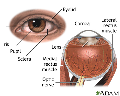
External Anatomy Of The Eye
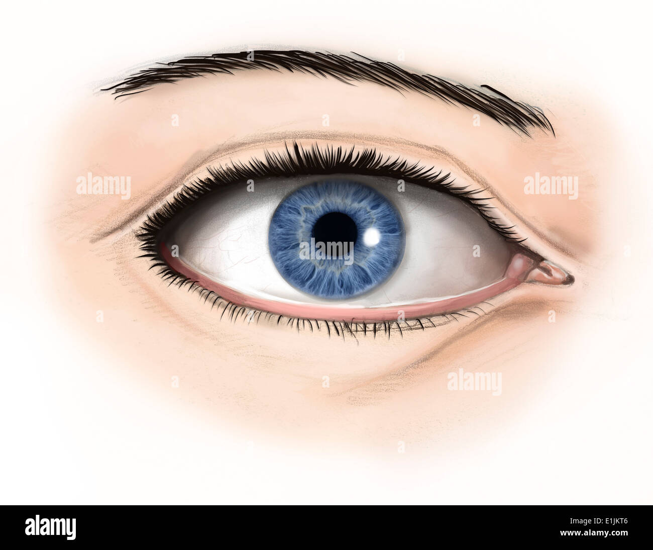
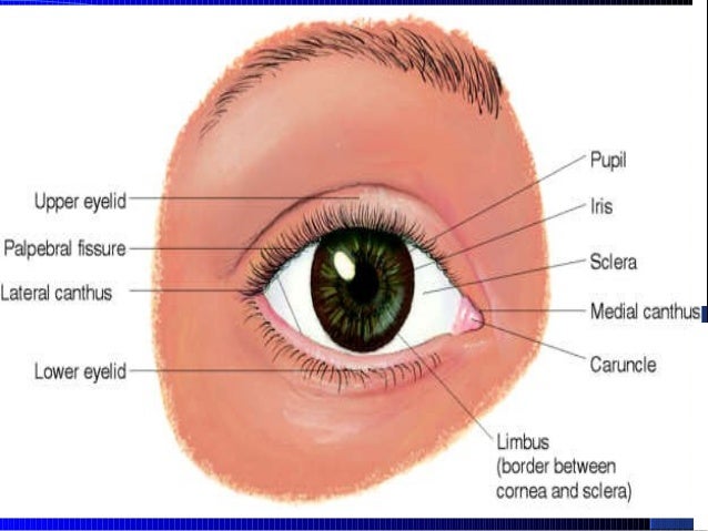
It is the most visible part of the eye. The outer margin of the cornea.
 External Eye Eye Drawing Tutorials Eye Anatomy Realistic
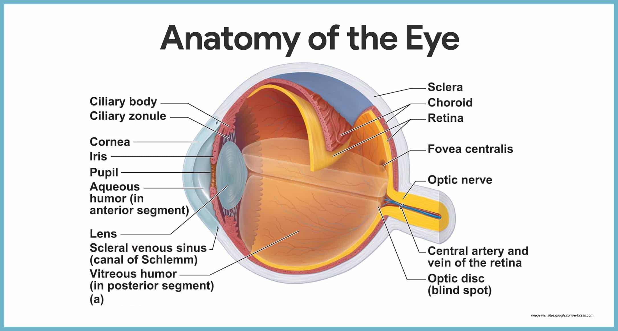
The human eye ball is spherical in structure and is about 24 mm in a diameter.

External anatomy of the eye. Component of the lacrimal apparatus tears enter thru lacrimal punctum. The episclera is covered by the conjunctiva a vascular mucous membrane. The eye is surrounded by the orbital bones and is cushioned by pads of fat within the orbital socket.
The lens then changes shape to allow the accurate focusing of light on the retina. Anatomy of the eye. The diaphragm that controls the amount of light entering the eye.
It has two layers a posterior pigment epithelium and an anterior stroma made of collagen muscle and pigment cells. The outer fibrous or sclera 2. Squinting which is closing the eye partially sheilds the eye from excessive light that can damage the internal structure such as the retina.
The iris is the colored part of the eye that controls the amount of light that enters into the eye. Overview the cornea allows light to enter the eye. The structure of the human eye is made of three layers.
It lies in front of the crystalline lens and separates the anterior chamber from the posterior chamber. Nerve signals that contain visual information are transmitted through the optic nerve to the brain. Human eye parts 1.
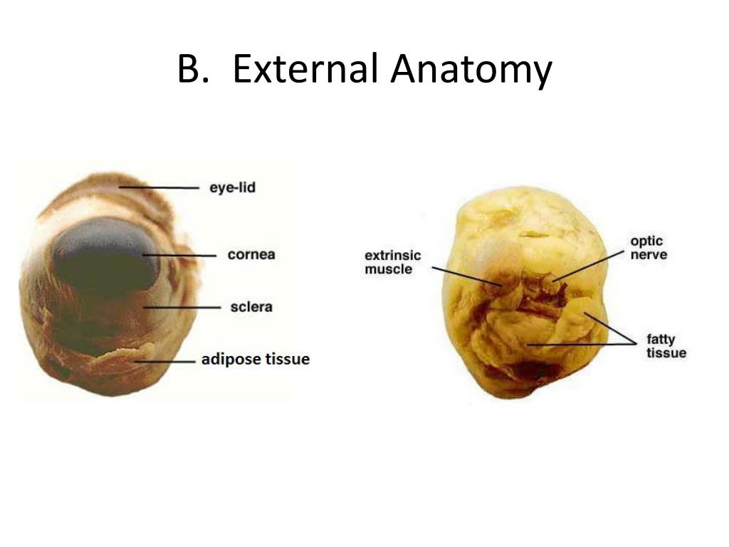
Extraocular muscles help move the eye in different directions. It is the most visible part of the eye. Malfunction in any part of the system can cause serious complications.
As light passes through the eye the iris changes shape by expanding and letting more light through or constricting and letting less light through to change pupil size. Blinking on the other hand which is closing and opening the eye rapidly spreads tears across and and removes irritants from the cornea and conjuctiva. Component of the lacrimal apparatus located superior and lateral to each eye.
Lacrimal system tear drainage system the lacrimal system is crucial for tear production and management which includes distribution of tears and draining excess tears.

 Anatomy Of The Eye
 External Anatomy Of The Eye And Accessory Structures Diagram
 The Anatomy Of The Human Eye Humaneyeproject
 External And Internal Eye Anatomy Medlineplus Medical

 The Eye And Vision
 Normal Eye Ultrasound How To

 External Anatomy Of The Human Eye With Labels Art Print By Art Com
 Eye Anatomy Labeling

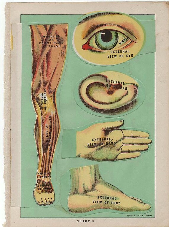 Five Overlays Of Eye Ear Hand Foot Leg 1916 Manikin Illustrations Of Internal And External Anatomy
 Special Senses Anatomy And Physiology Nurseslabs
 External Anatomy Of The Human Eye Stock Photo 69866838 Alamy
 Eyes
Eyes
 Eye Structure And Function In Cats Cat Owners Merck


 Ppt Sheep Eye Dissection Powerpoint Presentation Free
 Game Statistics External Anatomy Of The Eye Purposegames

 Understanding Glaucoma Anatomical Chart 9781587799280
 The External Anatomy Of The Eye Stock Image F002 4037
 External Anatomy Of Eye Diagram Quizlet
 Human Eye Wikipedia
 Eye Wikipedia
3 Eye External Anatomy Recorded Lecture Video Youtube

 External Anatomy Of The Human Eye Tote Bag
 Figure 23 1 External Anatomy Of The Eye And Accessory
 1 External Anatomy Of Eye A Frontal View B Side View
Belum ada Komentar untuk "External Anatomy Of The Eye"
Posting Komentar