Sural Nerve Anatomy
The sural nerve is a sensory nerve of the lower lateral leg and lateral aspect of the foot. Sural nerve formation at the distal third of the gastrocnemius both sural cutaneous branches join to become the sural nerve.
 Sural Nerve Png Images Sural Nerve Clipart Free Download
The sural nerve is a sensory nerve in the calf region of the leg.
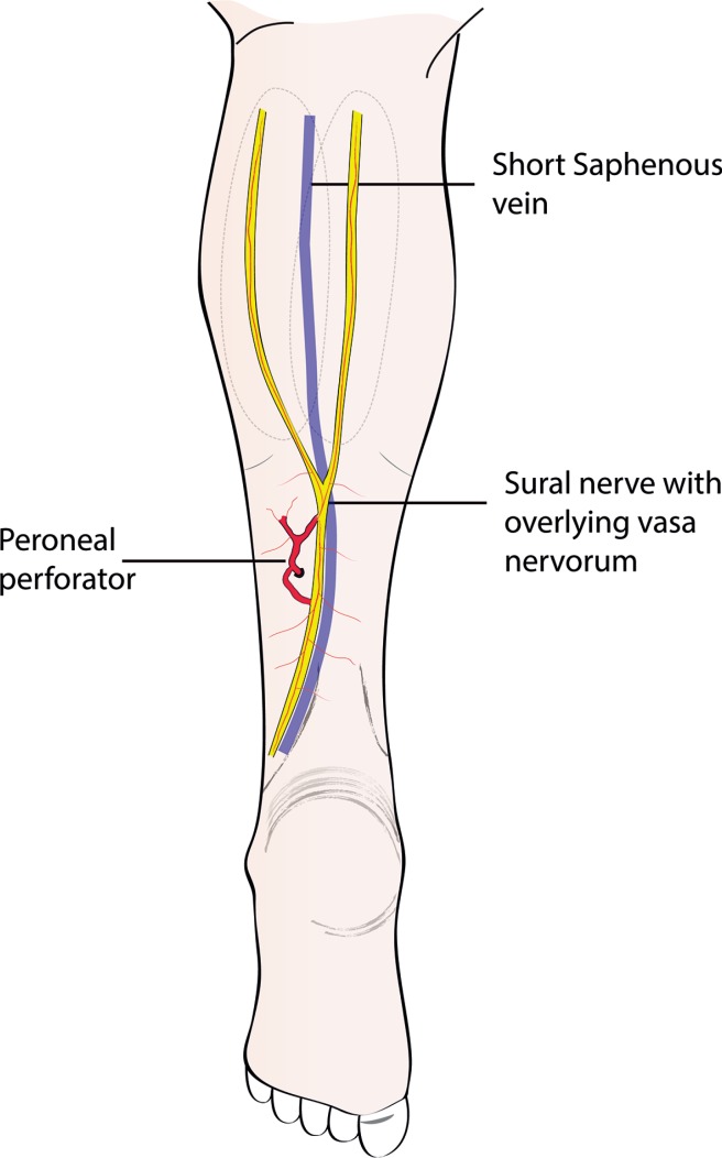
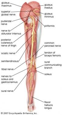
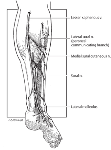
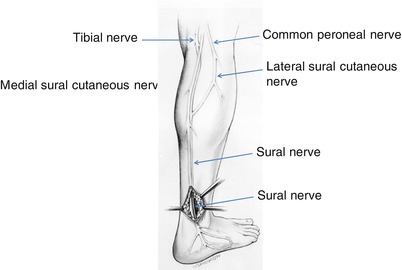
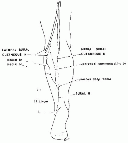
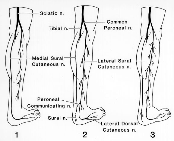
Sural nerve anatomy. High in the popliteal fossa the sciatic nerve divides into its two main branches on route to serve the leg namely the tibial nerve and the common fibular nerve. In the posterior calf the sural nerve emerges from between the two heads of the gastrocnemius muscle and runs with the small saphenous vein inferiorly to curve under the lateral malleolus. It can end at the lateral border of the foot without.
The short saphenous nerve initially courses posterior between the heads of the gastrocnemius muscle. Sural nerve anatomy as aforesaid it is purely a sensory nerve and does not consist of motor fibers. The nerve is comprised of spinal nerve roots from s1 and s2.
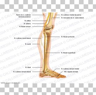
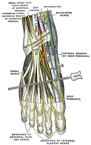
Descends on the posterolateral aspect of leg. Travels posterior to lateral malleolus and deep to fibularis tendon sheath. The sural nerve is a sensory nerve of the lower limb that supplies the lower posterolateral part of the leg and lateral part of the dorsum of the foot.
Clinical relevance damage to the sural nerve due to injury can occur as a result of trauma fractured calcaneus damage from surgery in the region. The sural nerve is a sensory nerve made up of collateral branches off of the common tibial and common fibular nerve. Functions the sural nerve supplies skin at the lower posterolateral side of the leg as well as the lateral aspect of the foot and little toe.
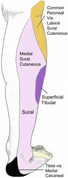
The sural nerve is purely sensory and it supplies sensation to the lower lateral leg lateral heel ankle and dorsal lateral foot. The sural nerve is a sensory nerve of the lower limb formed by the union of branches from the tibial nerve as well as common fibular nerve supplying sensation to the lower lateral aspect of the calf and foot. It travels within subcutaneous tissue adjacent to the small saphenous vein in the lower posterolateral calf.
The sural nerve supplies the dorsal cutaneous area of the lateral 2 and half toes in some cases. It is made up of branches of the tibial nerve and common fibular nerve the medial cutaneous branch from the tibial nerve and the lateral cutaneous branch from the common fibular nerve. It is generally described as a sensory nerve but may contain motor fibres discussed later in this article 14 16.
 Figure 3 From Late Estimation Of Sensibility Loss After
 Sciatic Nerve Decompression Background Indications
 Sural Nerve Anatomy Orthobullets
 Surgical Anatomy Of The Ovine Sural Nerve For Facial Nerve
 Lower Extremity Day 1 Flashcards Memorang
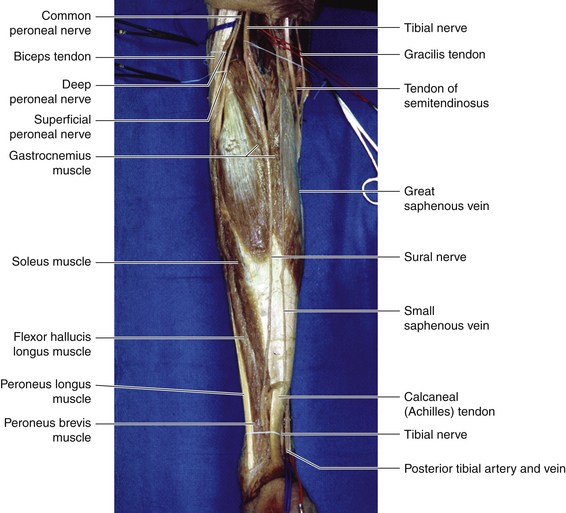
 The Tibial Nerve Course Motor Sensory Teachmeanatomy
 Sural Nerve Clinical Gate
 Tibial Nerve Radiology Reference Article Radiopaedia Org
 Sural Nerve Biopsy Learnneurosurgery Com
 Sural Nerve Injury And Neuroma Neupsy Key
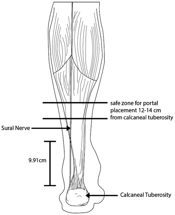 Cadaveric Anatomical Study Of Sural Nerve Where Is The Safe
 Sural Nerve Location Origin Function Nerve Block
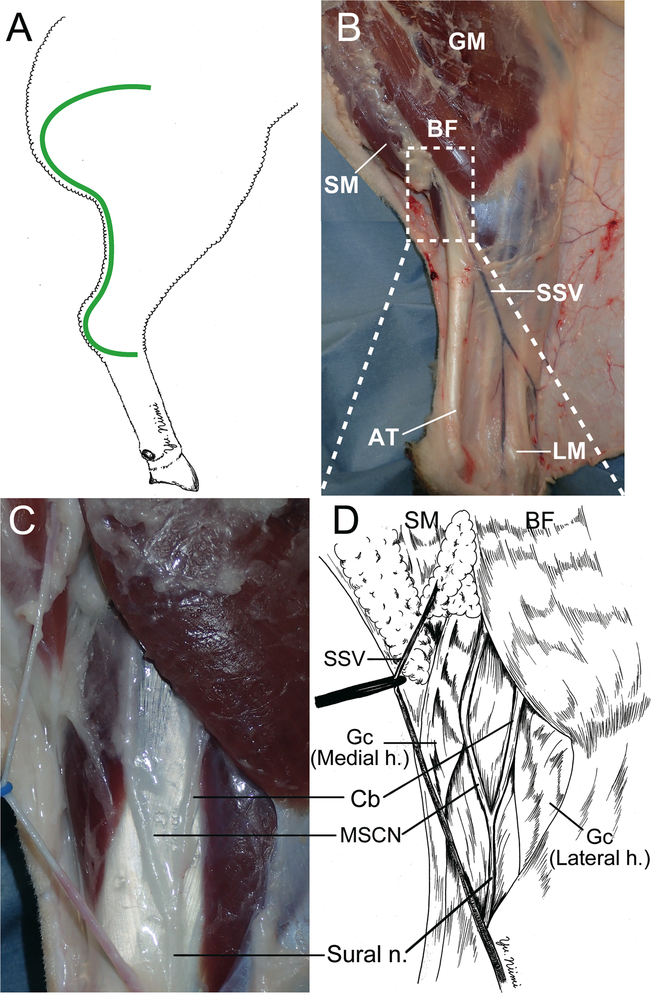
 The Surgical Anatomy Of Sural Nerve Sural N Middle A B
 Uncommon Injuries Sural Nerve Neuropathy
 Midfoot Approach Dorsolateral To The Cuboid Ao Surgery
 The Course Of The Sural Nerve And The Small Saphenous Vein
 Ankle Block Hadzic S Peripheral Nerve Blocks And Anatomy
![]() Common Peroneal Nerve Physiopedia
Common Peroneal Nerve Physiopedia
 Anatomical Variations Of The Formation And Course Of The
Distally Based Sural Artery Adipofascial Flap Based On A
 Stock Illustration
 Normal Anatomy Of The Peripheral Sural Nerve Springerlink
 Sural Nerve Entrapment Springerlink
 Nerves Musculoskeletal Key
 Anatomy Atlases Illustrated Encyclopedia Of Human Anatomic
Anatomy Bony Pelvis And Lower Limb Sural Nerve Article
 Cutaneous Nerve Blocks Of The Lower Extremity Nysora
 Nerve Biopsy Healthcare Baylor College Of Medicine
 Reconstruction St Louis Childrens Hospital
 Ankle Block
 A Porcine Model Of Peripheral Nerve Injury Enabling Ultra


Belum ada Komentar untuk "Sural Nerve Anatomy"
Posting Komentar