Sinus Anatomy
The ethmoid sinuses are located on each side of the bridge of your. Two types of sinus the blood filled and the air filled sinuses are discussed in this article.
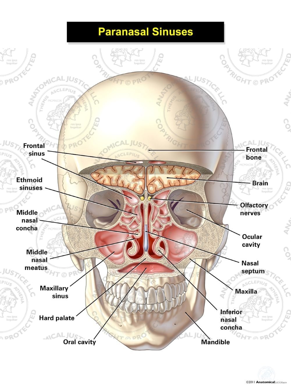 Paranasal Sinuses
The four paired sinuses or air cavities can be referred to as.

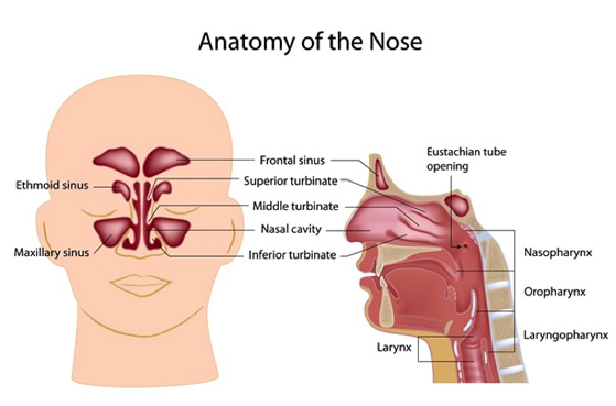
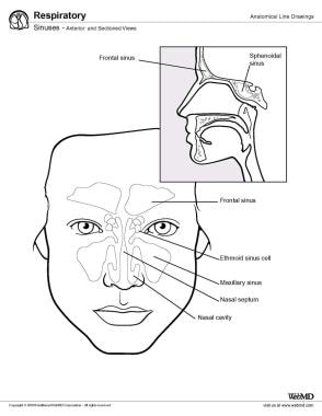
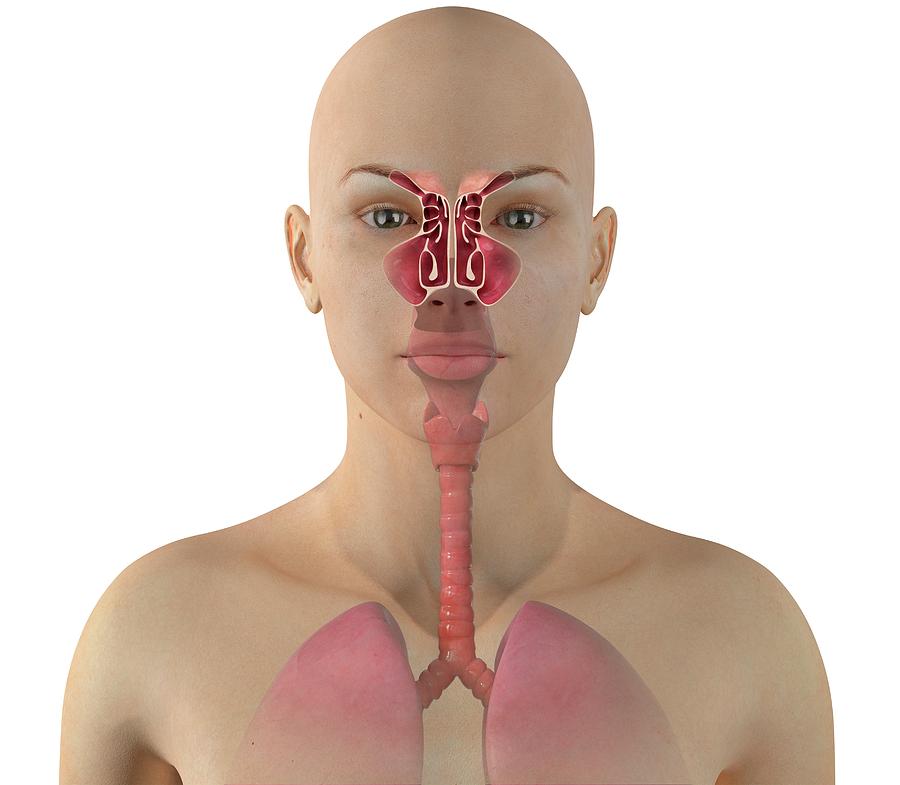
Sinus anatomy. Frontal sinus cavities which can be found above the eyes more in the forehead region. Maxillary sinus cavities are located on either side of the nostrils cheekbone areas. Between your eyes are your ethmoid sinuses.
Pilonidal sinus pilonidal cyst. Sinuses also have their own anatomy and function. Nasal sinuses are hollow air filled spaces within the bones of the skull and face.
Sinuses are found in our skull bones. Sinus anatomy the passages that begin with your nostril lead to a network of nasal passages and cavities in behind the face. Sphenoid sinus within the sphenoid bones.
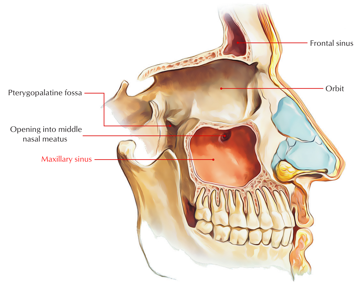
While you have a number of sinuses in your body those in your face which are known more technically as the paranasal sinuses are the most well known. The maxillary sinuses are located on each side of your nose near the cheek bones. The largest sinus cavities are about an inch across.
Sinus in anatomy a hollow cavity recess or pocket. Or a cavity within a bone. The sphenoid sinuses drain out onto the roof of the nasal cavity.
Definition of nasal sinuses. Ethmoid sinus cavities which are located between the eyes. These are known as sinuses.
Others are much smaller. The low center of your forehead is where your frontal sinuses are located. Many of us are not aware of our sinuses most of the time except when infection and other sinus disorders cause pain pressure sleep disorders or impaired hearing or breathing.
The sinuses are a connected system of hollow cavities in the skull. There are four pairs of sinuses named for the bones that theyre located in. Each sinus is named for the bone in which the sinus can be found.
Maxillary sinuses are the largest maxmaximum of the sinuses. There are three ethmoidal sinuses. A large channel containing blood.
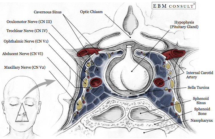
Your cheekbones hold your maxillary sinuses the largest. The relationships of this sinus are of clinical importance the pituitary gland can be surgically accessed via passing through the nasal roof into the sphenoid sinus and through the sphenoid bone. Anterior middle and posterior.
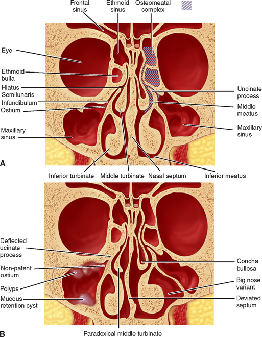
They are shaped like pyramids. The nasal septum separates the left and right maxillary sinuses 9. They empty into the nasal cavity at different places.
The frontal sinuses are located above the eyes near your forehead. Tentorial sinus straight sinus. Dermal sinus a congenital sinus tract extending from the surface of the body between the bodies of two adjacent lumbar vertebrae to the spinal canal.
The most posterior of all the paranasal sinuses these are located behind the ethmoid sinuses near the pituitary gland and optic nerves 8.
 38 Maxillary Sinus Anatomy Pathology And Graft Surgery
 Sinus Anatomy
 Dr Reuben Setliff Pioneer In Sinus Care
 Maxillary Sinus Earth S Lab
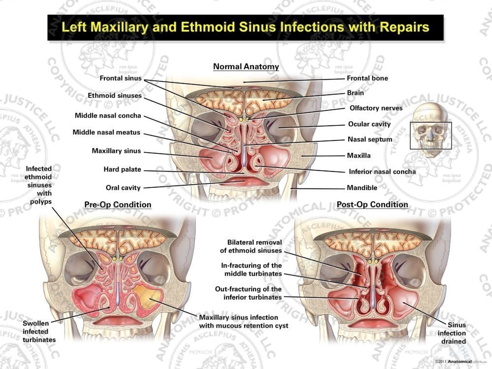 Left Maxillary And Ethmoid Sinus Infections With Repairs
 Dural Venous Sinuses Wikipedia
 Paranasal Sinus Anatomy Overview Gross Anatomy
 Anatomy And Physiology Paranasal Sinuses
 Startradiology
 Anatomy Of The Cavernous Sinus Purposegames
 Sinus Infection Sinusitis Symptoms Signs Treatment
Normal Sinus Anatomy Medivisuals Medical Illustration
 Customized 3d Sinus Anatomy Model With Back Card Buy Sinus Model Sinus Anatomy Neck Model Product On Alibaba Com
 Murine Sinus Anatomy A Sagittal Section Of The Skull And
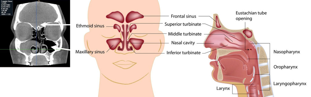
 Anatomy Of The Sinuses Otolaryngology Houston
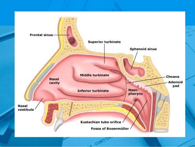
 Sinus Anatomy And Ct Scan Fort Worth Ent Sinus
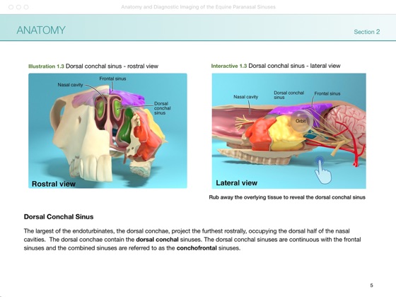 Anatomy And Diagnostic Imaging Of The Equine Paranasal
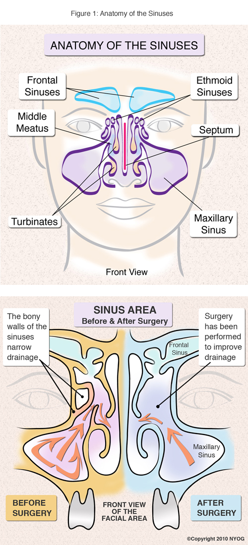 Sinus Anatomy Check Out This Helpful Diagram Ny Sinus Center
 Sinus Cancer Anatomy Headandneckcancerguide Org
 Sinus Anatomy 1
 Paranasal Sinuses An Overview Sciencedirect Topics
 Science Source Sinus Anatomy Illustration
 Sinusitis Allergy Asthma And Sinus Specialists City Allergy
Belum ada Komentar untuk "Sinus Anatomy"
Posting Komentar