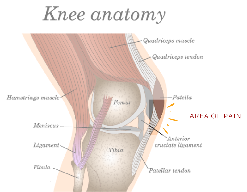
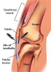
Patellar Tendonitis Anatomy
The patellar ligament is usually around 5 cm in length but its length. The patellar tendon arises from the apex of the patella as well as its anterior.
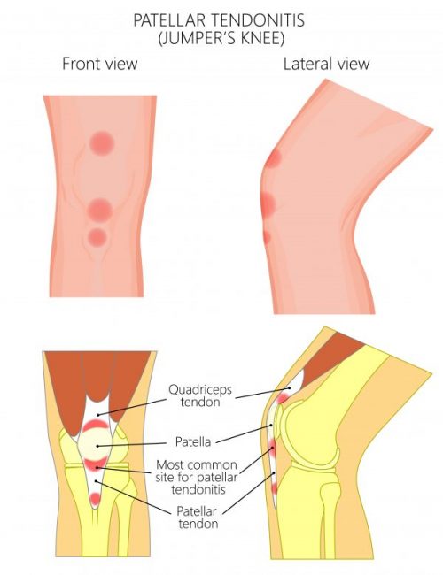
This occurs mostly in athletes from repeated jumping.
Patellar tendonitis anatomy. Summary the patella is a sesamoid bone that lies in the tendon of quadriceps femoris. Once the point of maximal tenderness is identified the knee is flexed to 90 and pressure is again applied to the tendon figure 1b. Where you will feel the pain.
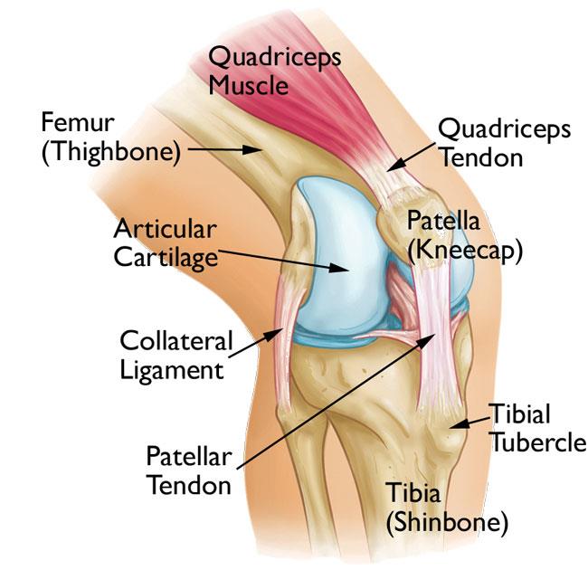
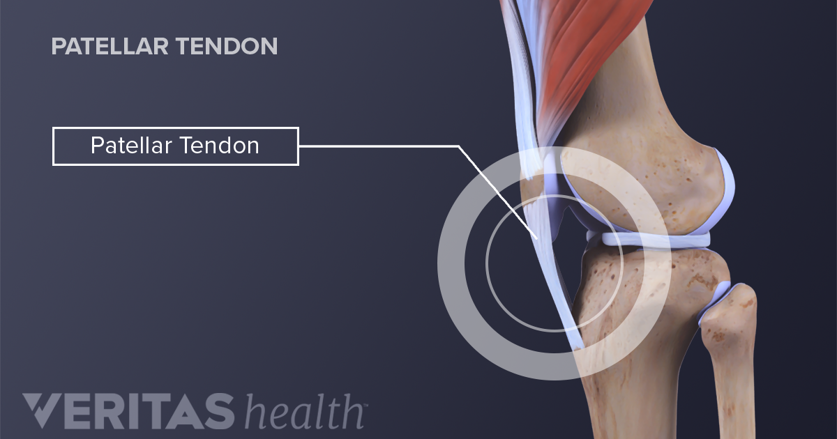
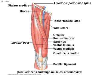
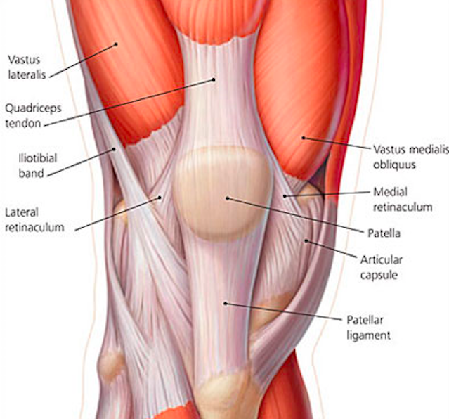
Functional anatomy the rectus femoris and 3 vasti muscles ie the vastus medialis vastus lateralis and vastus intermedius muscles join in a common quadriceps tendon that inserts on the patella. The quadriceps tendon connects the quadriceps muscles of the thigh to the kneecap and provides the power for straightening the knee. The largest tendon in the knee is the patellar tendon which covers the kneecap runs up the thigh and attaches to the quadriceps.
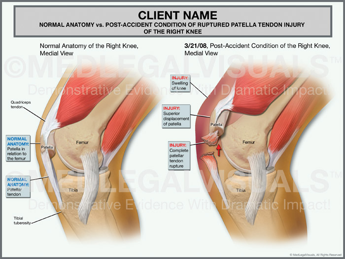
In the case of patellar tendinitis tenderness to palpation of the tendon is most often located at the origin of the tendon at the inferior pole of the patella. Three bones meet to form your knee joint. Patellar tendinopathy is primarily.
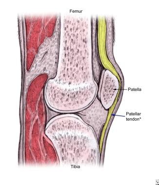
The patellar tendon inserts onto the tibial tuberosity. Inflammation of the tendon connecting the kneecap patella to the shin bone. There are two major tendons in the kneethe quadriceps and patellar.
Bones cartilage ligaments and tendons. The patellar tendon connects your patella the kneecap to the shinbone. Tendons in the knee.
It is made up of four main things. Patellar tendinopathy is a source of anterior knee pain characterised by pain localised to the inferior pole of the patella. This tendon can withstand very high forces but in spite of its durability the patellar tendon can still wear down over time if its frequently overstressed.
This occurs mostly in athletes from. Pain is aggravated by loading and increased with the demand on the knee extensor musculature notably in activities that store and release energy in the patellar tendon. It also helps hold the patella in the patellofemoral groove in the femur.
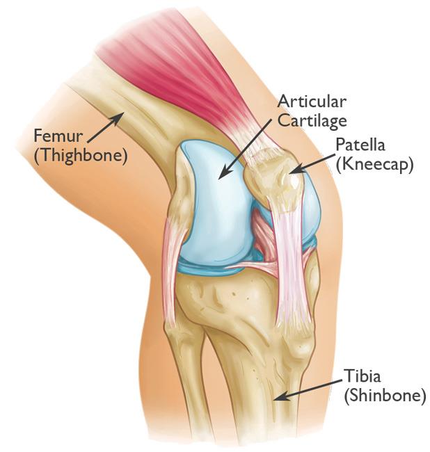
You use this tendon every time you straighten your knee. Your thighbone femur shinbone tibia and kneecap patella. The ends of the femur and tibia and the back of the patella are covered with articular cartilage.
 Patellofemoral Pain Syndrome Orthoinfo Aaos
Injuries To The Knee Ii March April
 Patellar Tendonitis Exercises Treatment Enerskin
 Patellar Tendinopathy Irritation Of The Patellar Tendon

 Patellar Ligament Wikipedia
 Evolution Of The Patellar Sesamoid Bone In Mammals Peerj
 How To Heal From A Ruptured Patellar Tendon Injury Premier
 Collateral Ligaments Of The Knee Joint Patellar Tendon
Prepatellar Kneecap Bursitis Orthoinfo Aaos

 Symptoms Of An Acute Patellar Injury
 Patellar Tendon Pain Squat University
 Quadriceps Tendon Tear Physiopedia
 Patellar Tendonitis Jumper S Knee Central Coast
![]() Patellar Tendon Anatomy Origin Insertion Function Kenhub
Patellar Tendon Anatomy Origin Insertion Function Kenhub
 Patellar Fractures Broken Kneecap Orthoinfo Aaos
 Jumper S Knee Symptoms In Kids Jumper S Knee Treatment
 Quadriceps Tendon Rupture Core Em
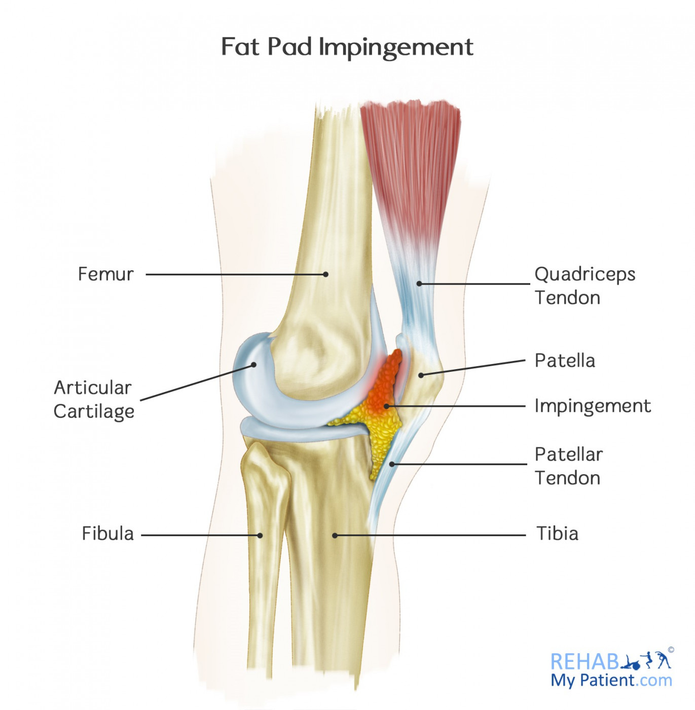 Fat Pad Impingement Rehab My Patient
 Patella Wikipedia

 Fat Facts For Clinicians Treating Anterior Knee Pain
 Jumper S Knee Background Epidemiology Functional Anatomy
![]() Patellar Tendon Anatomy Origin Insertion Function Kenhub
Patellar Tendon Anatomy Origin Insertion Function Kenhub
 Patellar Tendonitis How To Get Rid Of Jumper S Knee
 Jump Mechanics And Risk Of Patellar Tendinopathy Lower
 Summit Medical Group
Belum ada Komentar untuk "Patellar Tendonitis Anatomy"
Posting Komentar