Hip Anatomy Mri
Click on a link to get t1 axial view t1 coronal view. About anatomy mri magnetic resonance imaging is particularly well suited for the medical evaluation of the musculoskeletal msk system including the knee shoulder ankle wrist and elbow.
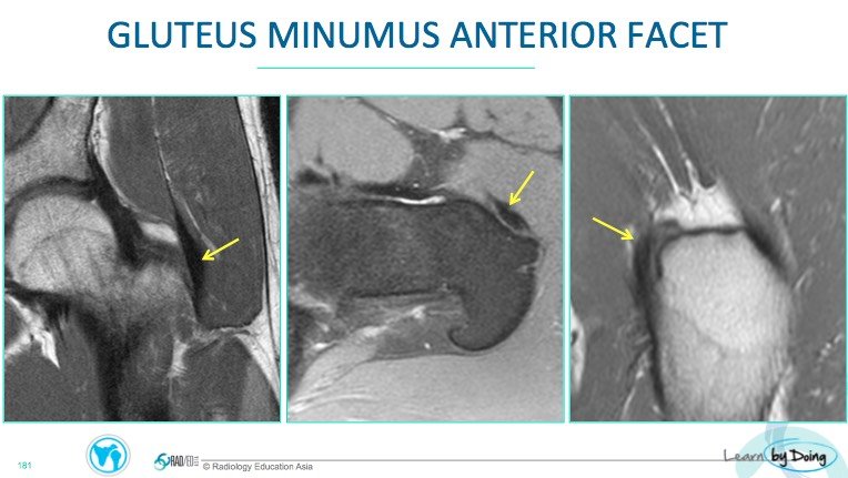 Mri Hip Gluteal Tendon Anatomy Radedasia
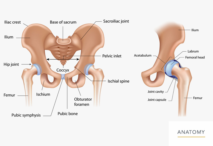
The hip joint is a synovial joint between the femoral head and the acetabulum of the pelvis.

Hip anatomy mri. Since this joint transfers weight from the upper body to the lower limbs it is subject to a range of problems resulting from faulty weight bearing positions in normal individuals to problems caused by wear and tear in those who are athletically active. Use the mouse scroll wheel to move the images up and down alternatively use the tiny arrows on both side of the image to move the images. This webpage presents the anatomical structures found on hip mri.
This mri hip joint coronal cross sectional anatomy tool is absolutely free to use. Injuries such as anterior cruciate ligament meniscus and rotator cuff tears are all easily diagnosed when there is a firm understanding and knowledge of human anatomy. This article considers the hip joint specifically however it is worth noting that the word hip is often used to refer more generally to the anatomical region around this joint.
Mri of the hip. Use the mouse scroll wheel to move the images up and down alternatively use the tiny arrows on both side of the image to move the images. Stanford bone tumor bayesian network issssr msk lectures for residents ocad msk cases from around the world stanford msk mri atlas has served almost 800000 pages to users in over 100 countries.
Knee shoulder shoulder arthrogram ankle elbow wrist hip contact. Mri of the hip joint the hip joint is a ball and socket type of joint that is also the deepest joint in the body. Use the mouse to scroll or the arrows.
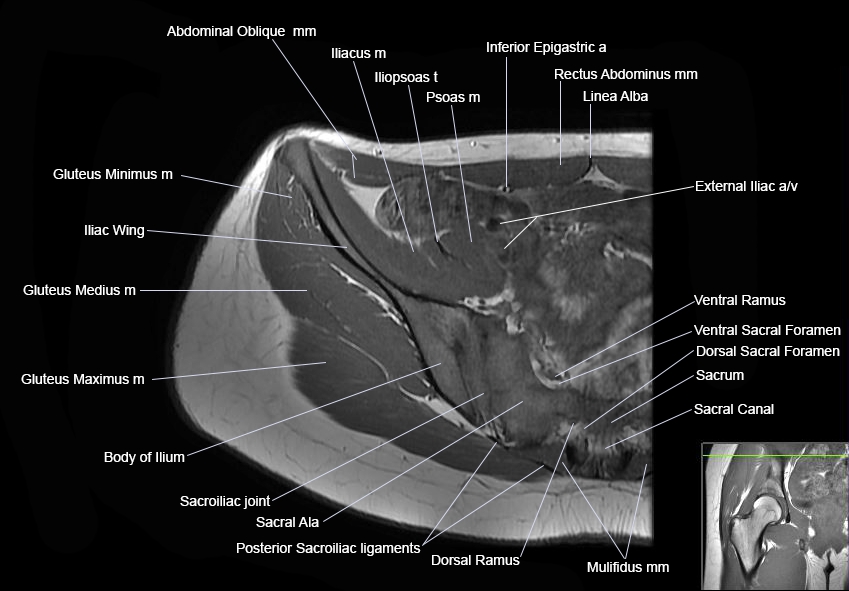
This mri hip joint axial cross sectional anatomy tool is absolutely free to use. The hip anatomy on 3t mr and 3d pictures on these 252 3t mri images over 340 anatomical structures are captioned. At the end of the module there are 3d reconstructions of the hip joint hip bone and femur as a recapitulation of musculoskeletal anatomy.

 Anatomy Of Hip Joint Free Mri Coronal Cross Sectional
 Imaging The Hip Musculoskeletal Imaging Handbook F A
 Mri Hip Anatomy Www Unidadortopedia Com Pbx 6923370
 Amazon Com Volume Ii Mri Of Sports Hernias Thigh And Hip
 Mri Anatomy Of Hip Joint Free Mri Axial Hip Anatomy

Module 2 Lower Extremity Orthopedic Imaging

 Ecr 2016 C 0492 Imaging Findings Of Developmental
 K Anatomy
 Hips Positioning And Mri Anatomy El Paso Back Clinic

 Mri Hip Jiont Anatomy Dr Ahmed Eisawy
 Joshua Harris Md On Twitter Hip Flexor Problems
 Imaging Of The Hip And Pelvis Radiology Key
 Hip Mri
 The Hip Anatomy On 3t Mr And 3d Pictures
Hip Bursitis Orthoinfo Aaos
 Hip Joint
 Anatomy Of Hip Joint Free Mri Coronal Cross Sectional
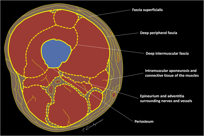 Fasciae Of The Musculoskeletal System Mri Findings In
 Hip Mri Planning Procedure Radtechonduty
 Diagnostic Imaging Of The Hip For Physical Therapists
![]() Medical Imaging And Radiological Anatomy X Ray Ct Mri
Medical Imaging And Radiological Anatomy X Ray Ct Mri
Osteonecrosis Of The Hip Orthoinfo Aaos
 Hip Thigh Orthopedic Specialist Of Northern California
 Mri Anatomy Of Hip Joint Free Mri Axial Hip Anatomy
 Hip Mri
Belum ada Komentar untuk "Hip Anatomy Mri"
Posting Komentar