Heart Structure Anatomy
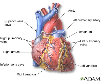
The auricles of the atria are visible on the left side surrounding the root of the aorta and the pulmonary trunk whilst the large veins and the main parts of the atria are situated on the right. The heart is enclosed in a pericardial sac that is lined with the parietal layers of a serous membrane.
 Heart Anatomy Anatomy And Physiology
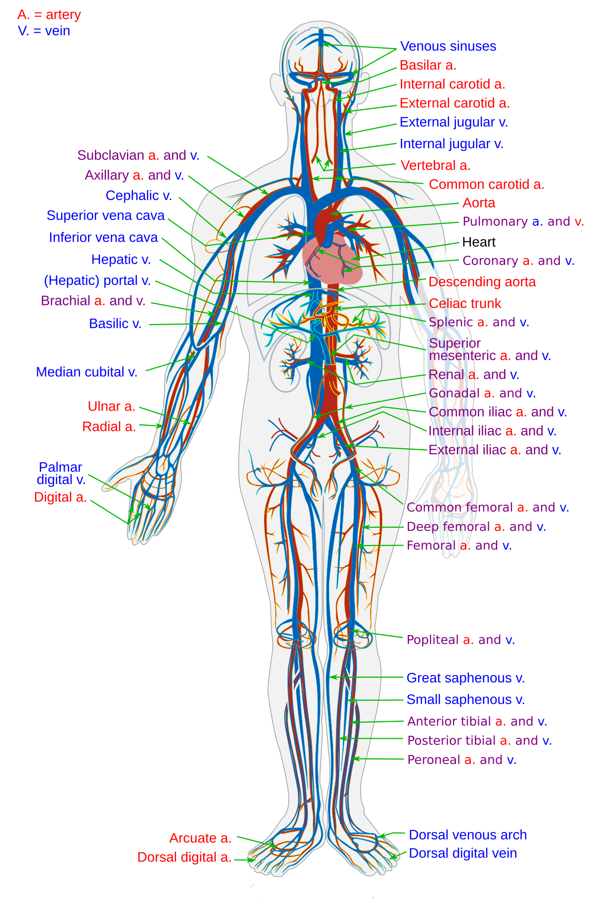
The circulatory system lower body image with blank labels attached.
Heart structure anatomy. Images and pdfs. 12 cm 5 in in length 8 cm 35 in wide and 6 cm 25 in in thickness. Shape and size of the heart.
The heart an image of the heart with blank labels attached. Grooves on the surface represent the divisions of the internal structure of the heart. Structure of the heart the human heart is a four chambered muscular organ shaped and sized roughly like a mans closed fist with two thirds of the mass to the left of midline.
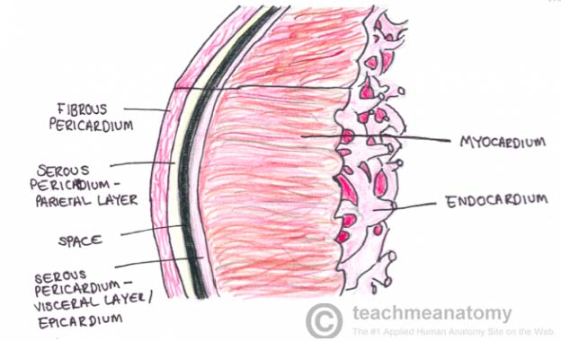
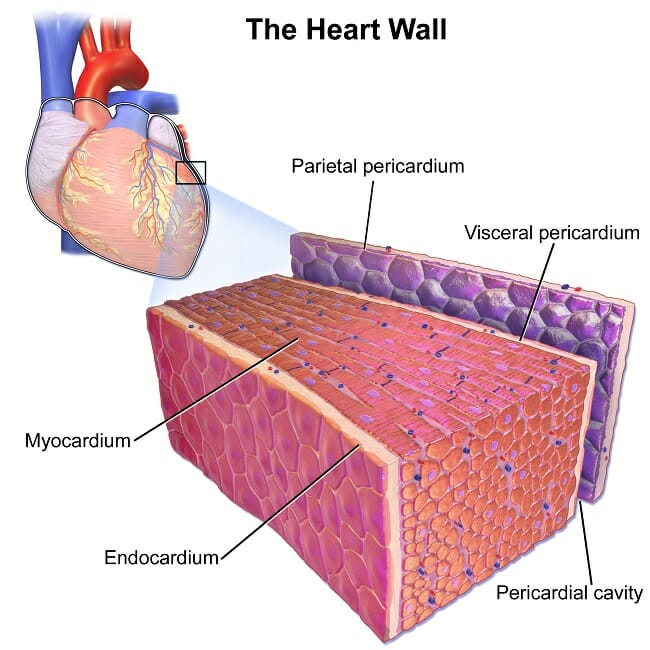
A typical heart is approximately the size of your fist. The heart is situated within the chest cavity and surrounded by a fluid filled sac called the pericardium. Layers of the heart wall.
The heart is a muscular organ about the size of a fist located just behind and slightly left of the breastbone. Human heart gross structure and anatomy heart. Position and shape of the heart.
The circulatory system upper body image with blank labels attached. Here we will review its essential components and how and why blood passes through them. The heart pumps blood through the network of arteries and veins called the cardiovascular system.
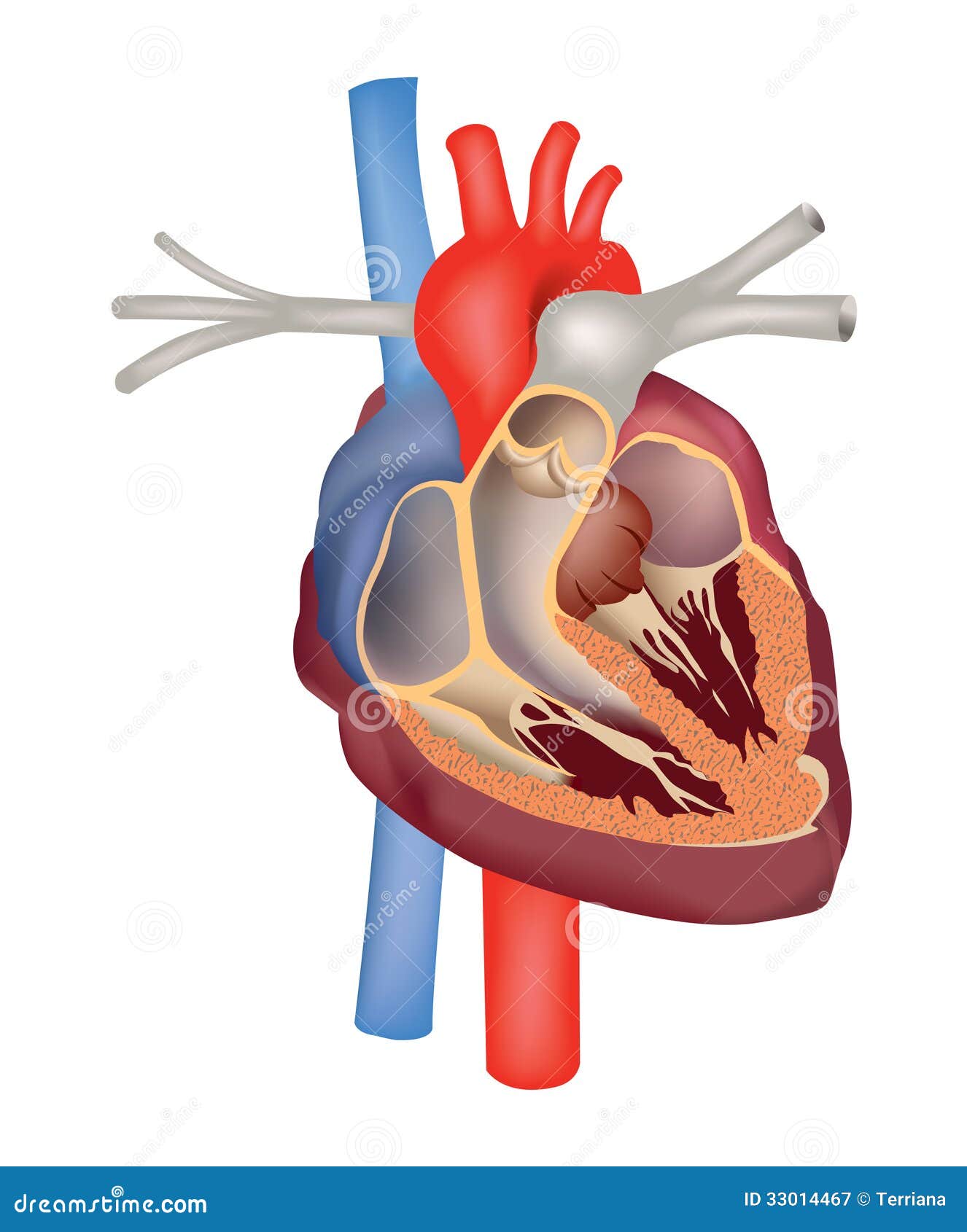
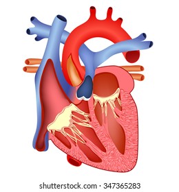
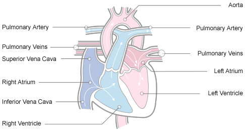
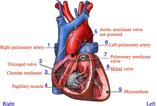
The anatomy of the heart. The circulatory system a pdf file of the upper and lower body for printing out to use off line. The chambers of the heart are shown including the right atrium left atrium right ventricle and left ventricle.
This 3d medical animation depicts the anatomy of the heart. The right atrium receives blood from the veins and pumps it to the right ventricle. This amazing muscle produces electrical impulses that cause the heart to contract.
The heart has three layers of tissue each of which serve a slightly different purpose. Chambers of the heart. The hearts unique design allows it to accomplish the incredible task of circulating blood through the human body.
The heart has four chambers. The shape of the heart is similar to a pinecone rather broad at the superior surface and tapering to the apex. Heart is a myogenic muscular organ that pumps blood throughout the blood vessel by repeated rhythemic contraction.
It is divided by a partition or septum into two halves and the halves are in turn divided into four chambers.
 Cardiac Muscle Definition Function And Structure
 Human Heart Muscle Structure Anatomy Diagram Clip Art
 Heart Structure Anatomy Heart Cross Section Stock Vector
 Heart Structure Images Stock Photos Vectors Shutterstock
 Anatomy Structure Of Heart In Hindi Bhushan Science
 Anatomy And Physiology Of The Heart Normal Function Of The
 How Your Heart Works Wake Forest Baptist Health
 The Human Heart Structure 3 Editable Power Point Slides Stem
 Heart Anatomy Anatomy And Physiology
 Human Heart Muscle Structure Anatomy Infographic Chart Diagram
 The Human Heart External And Internal Structure Online
 The Heart Structure And Function
 Section 2 Heart Structure Function At Grand Valley State
 How To Draw The Internal Structure Of The Heart 13 Steps
 Heart Structure Diagram Quizlet
 Anatomical Structure Of The Heart Cardiology An
 Human Heart Muscle Structure Anatomy Diagram Art Print Poster
 Studying Human Heart Diagram Heart Diagram Heart Structure
Essay On Human Heart Location Structure And Other Details
 Heart Structure Diagram Quizlet
 Congenital Heart Defect Wikipedia
 Illustration Of External Human Heart With Their Parts
 Free Art Print Of Human Heart Muscle Structure Anatomy Diagram
Mammalian Heart And Blood Vessels Boundless Biology

Belum ada Komentar untuk "Heart Structure Anatomy"
Posting Komentar