Finger Anatomy Bones
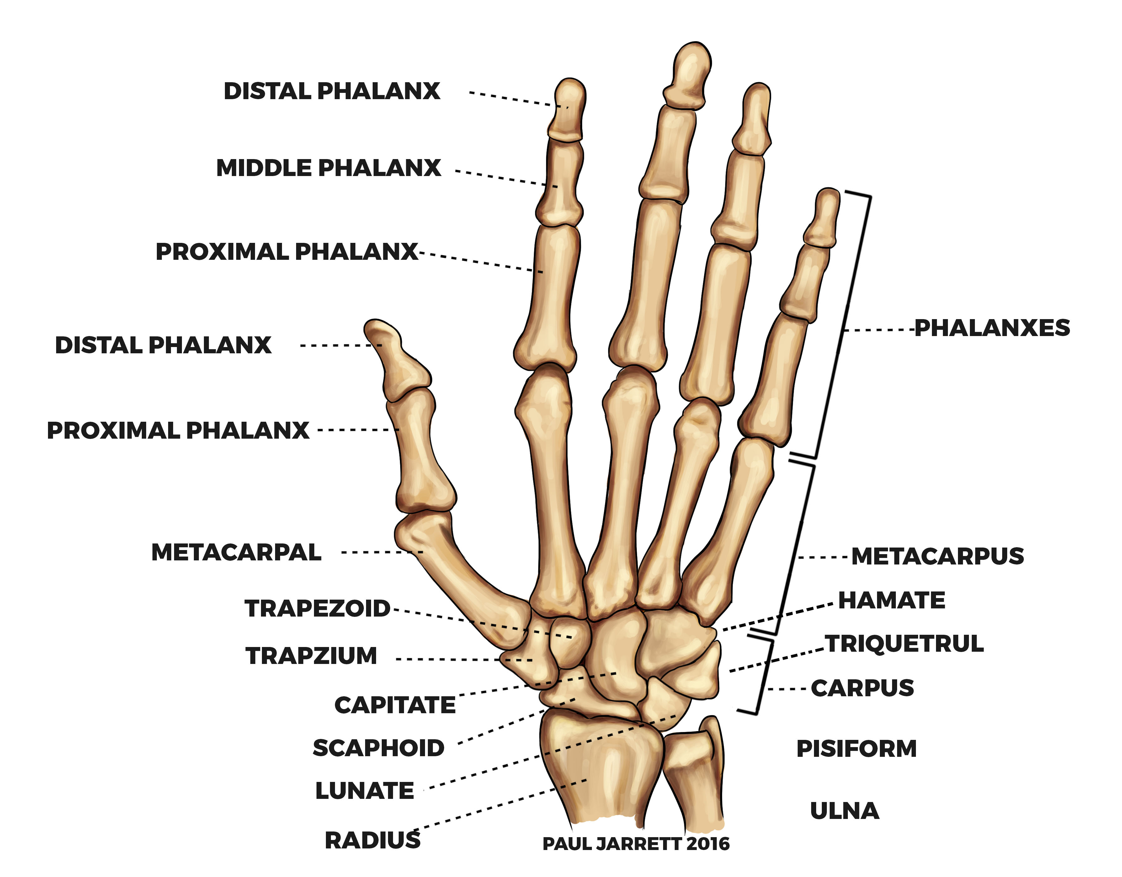
The finger bones are known as phalanges singular phalanx. They give the hand its structure and serve as an attachment for many small muscles tendons and ligaments in the hand.
5 elongated metacarpal bones which are next to the wrist and help to make up the palm.

Finger anatomy bones. Carpal bones proximal a set of eight irregularly shaped bones. Phalanges distal the bones of the fingers. A proximal phalanx articulates with the first metacarpal and a distal phalanx forms the tip of the thumb.
There are two interphalangeal joints ip joints on each finger. Anatomy of the fingers finger bones. Nerves send signals from the brain to the.
We use our hands in performing so many minor as well as major. The thumb only consists of a proximal and distal phalanx. These hand bones are five in number with each one of them relating to some digit.
Together with the phalanges of the fingers and thumb these metacarpal bones form five rays or poly articulated chains. Each thumb has two phalanges. Besides all scaphoid is the most commonly fractured bone in the hand.
The hand is made up of many bones. Finger bones are characterized by how near or far they are from the rest of the body. Each finger is made up of 3 phalanges.
The four fingers each consist of three phalanx bones. Each finger has three phalanges. There are no muscles in the fingers.
The metacarpals of the fingers make up the bone structure of most of the hand. Nerves of the fingers. Hand bones hand bones anatomy and structure.
Ask a doctor online now. Proximal middle and distal phalanges. 14 phalanges which make up the fingers.
The thumb is made up of 2. The final bone which is smallest and furthest from the hand is called the distal phalanx. The phalanges are fairly simple bones.
These 19 bones collectively form 14 separate joints. The subsequent bone next to the proximal phalange is the middle phalanx. The index and middle finger metacarpals have very little motion while the metacarpals of the ring and little finger move much more.
Proximal middle and distal. Finger movement is controlled by muscles in the forearms that pull on finger tendons. They are all similar in shape and have joints in the wrist on one end and the finger at the other end.
Metacarpals there are five metacarpals each one related to a digit. The rest of the fingers have three phalanges each. And fingers move by the pull of forearm muscles on the tendons.
The finger and thumb bones are called the phalanges. Fingers are constructed of ligaments strong supportive tissue connecting bone to bone tendons attachment tissue from muscle to bone and three phalanges bones. These are located in the wrist area.
Metacarpals are hand bones that line up with the fingers. They can be divided into three categories. The bone closest to the palm is called the proximal phalanx.
 A Digital Post Fingers And Thumbs
 Human Being Anatomy Skeleton Hand Image Visual
 Real Human Finger Bones Metacarpal Natural Bone
 Bones Of The Human Hand My Poor Right 3rd Distal Phalange
 8 2 Bones Of The Upper Limb Anatomy And Physiology
 Hand Finger Bone Diagram Hand Structure Anatomy Hand Finger
 Clinical Anatomy Hand Wrist Palmar Aspect Flexors
 Human Hand Parts Bones Left Hand Stock Illustration 620794583
 Bones And Joints Of A Human Hand Hand Anatomy Bone Joint
 Wrist Bones Kirkland Wa Evergreenhealth
 Carpal Tunnel Syndrome Symptoms And Treatment Orthoinfo
 Finger Anatomy Bones Joints Muscle Movements And Nerves
 Hand And Wrist Anatomy Murdoch Orthopaedic Clinic
 Bones Of The Hand Carpals Metacarpals Phalanges
 Anatomy 101 Finger Bones The Handcare Blog
 Phalange Bone An Overview Sciencedirect Topics
 Anatomy Of Hand Wrist Bones Muscles Tendons Nerves
 Hand Anatomy Midwest Bone Joint Institute Elgin Illinois


Belum ada Komentar untuk "Finger Anatomy Bones"
Posting Komentar