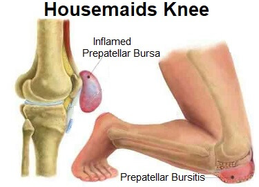
Knee Anatomy Bursa
The knee contains three important groups of bursae. Between the patellar ligament and the anterior part of the tibia bursitis of the knee is an inflammation of any of those eight bursae resulting in pain and tenderness of the knee swelling and a warm feeling when you touch the area.
 Foot Sinew Knee Anatomy Lateral Malleolus Retrocalcaneal
There are bursa located underneath the tendons and ligaments on both the lateral and medial sides of the knee.

Knee anatomy bursa. The prepatellar bursae lie in front of the patella. When one becomes inflamed increased tension and pain can occur in a temporary condition known as bursitis. The prepatellar bursa is one of the larger bursae of the knee and is located on the front of the patella hence pre patellar just under the skin.
A knee bursa basically functions as a cushion. It protects the patella. A knee bursa also known as a subcutaneous prepatellar bursa aids with movement when we walk run stretch or even cross our legs.
Knee bursitis is inflammation of a small fluid filled sac bursa situated near your knee joint. A bursa is a fluid filled structure that is present between the skin and tendon or tendon and bone. Sagittal section of right knee joint thus showing only frontal bursae.
Anatomy of the knee bursae a bursa is a small sac made of fibrous tissue that has an inner lining of synovial type membrane. Knee bursitis causes pain and can limit your mobility. Here we will look at knee bursa anatomy where they are found how they are injured and how to treat knee bursitis bursa are found all over the body and there are approximately fourteen around the knee.
The bursae can become irritated by frequent kneeling. The bursae of the knee are the fluid sacs and synovial pockets that surround and sometimes communicate with the joint cavity. Between the femur and quadriceps femoris it is attached to the articularis genu muscle and communicates with the synovial cavity.
It is filled with synovial fluid or lubricant made by the membrane. Between the skin and patella. Knee bursa are small fluid filled sacs which contain synovial fluid.
Typically bursae are located around large joints such as the shoulder knee hip and elbow1 inflammation of this fluid filled structure is called bursitis. Between the skin and tibial tuberosity. They sit between two surfaces usually muscle and bone.
There are four bursae anterior to the knee joint. Bursae one is a bursa are fluid filled sacs that help cushion the knee. Thin walled and filled with synovial fluid they represent the weak point of the joint but also produce enlargements to the joint space.
The main function of a bursa is to reduce friction between adjacent moving structures.
 Knee Pain On The Inside Of Your Joint Causes Solutions
 Anatomy Of The Knee Joint Knee Joint Anatomy Bursitis
 Knee Joint Ligaments And Bursae
 Infrapatellar Bursitis Or Clergyman S Knee Symptoms
 Prepatellar Kneecap Bursitis Orthoinfo Aaos
 Housemaids Knee Symptoms Diagnosis Treatment
 Knee Pain And Problems Loma Linda University Health
 Prepatellar Bursitis Physiopedia
 Prepatellar Bursa Musculoskeletal Anatomyzone
 Knee Pain Medlineplus Medical Encyclopedia Image
 Knee Bursae Locations Anatomy Study Com
 What Causes A Swollen Knee Water On The Knee
 Impingement Syndrome Brisbane Knee And Shoulder
 Articular Capsule Of The Knee Joint Wikipedia
 Knee Bursa Anatomy Function Injuries Knee Pain Explained
 Articular Capsule Of The Knee Joint Wikipedia
 Housemaids Knee Prepatellar Bursitis Information Sinew
 Knee Bursae Anatomy Diagram Quizlet
 Knee Pain On The Inside Of Your Joint Causes Solutions
 Bursitis Of The Knee Healthlink Bc
 Iliopsoas Bursitis Physiopedia
 Patient Education Concord Orthopaedics
 Shoulder Bursitis Pain Symptoms Treatment Pictures


Belum ada Komentar untuk "Knee Anatomy Bursa"
Posting Komentar