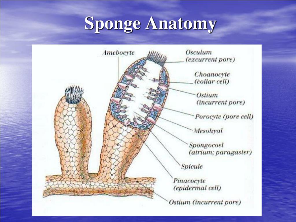
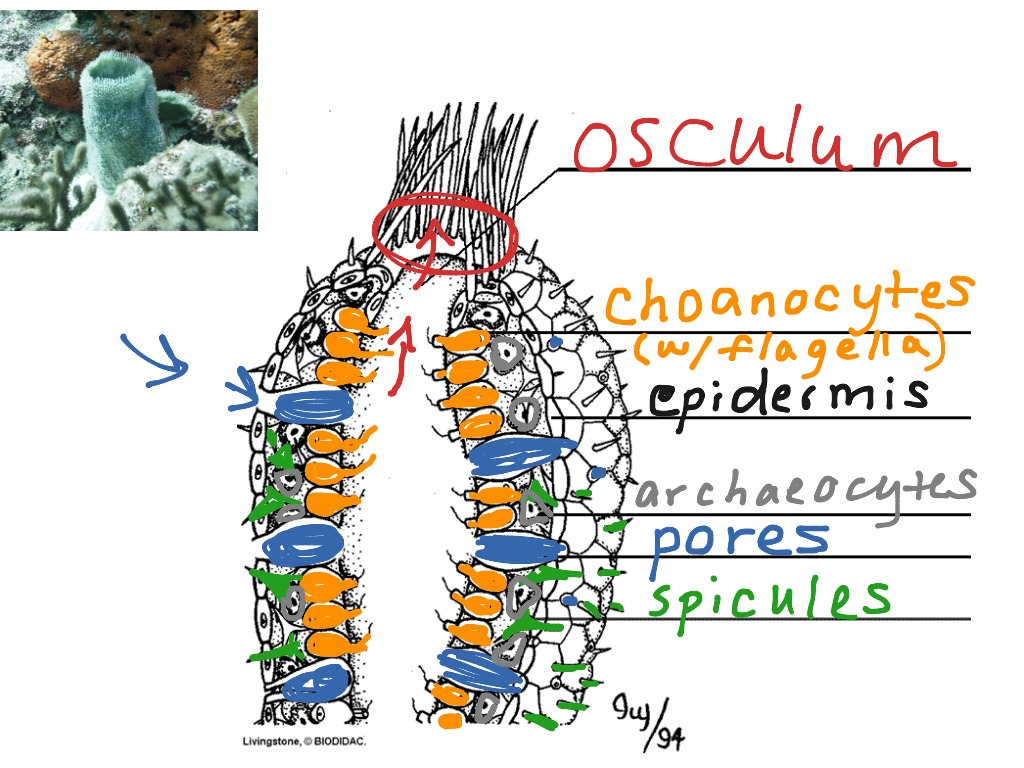
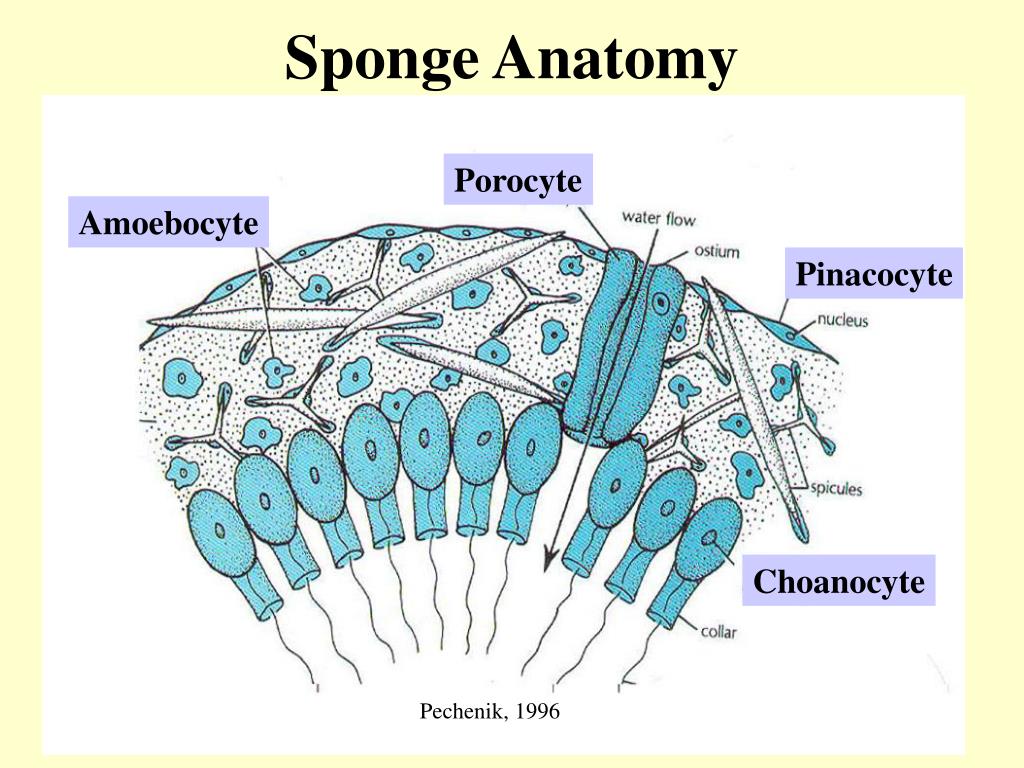
Sponge Anatomy
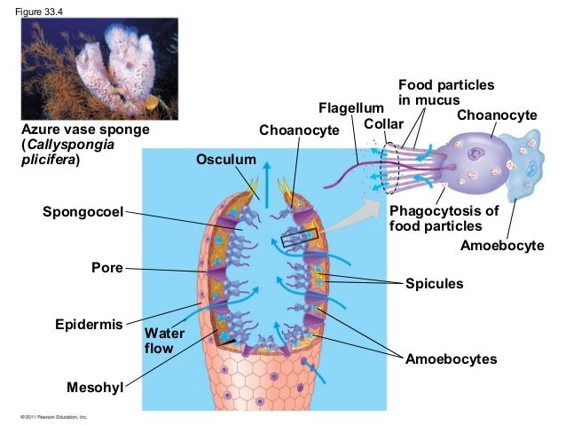
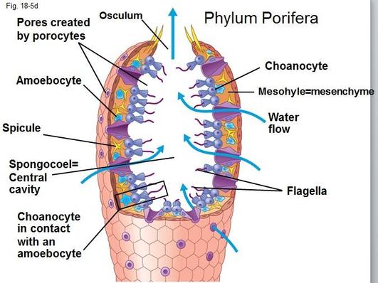
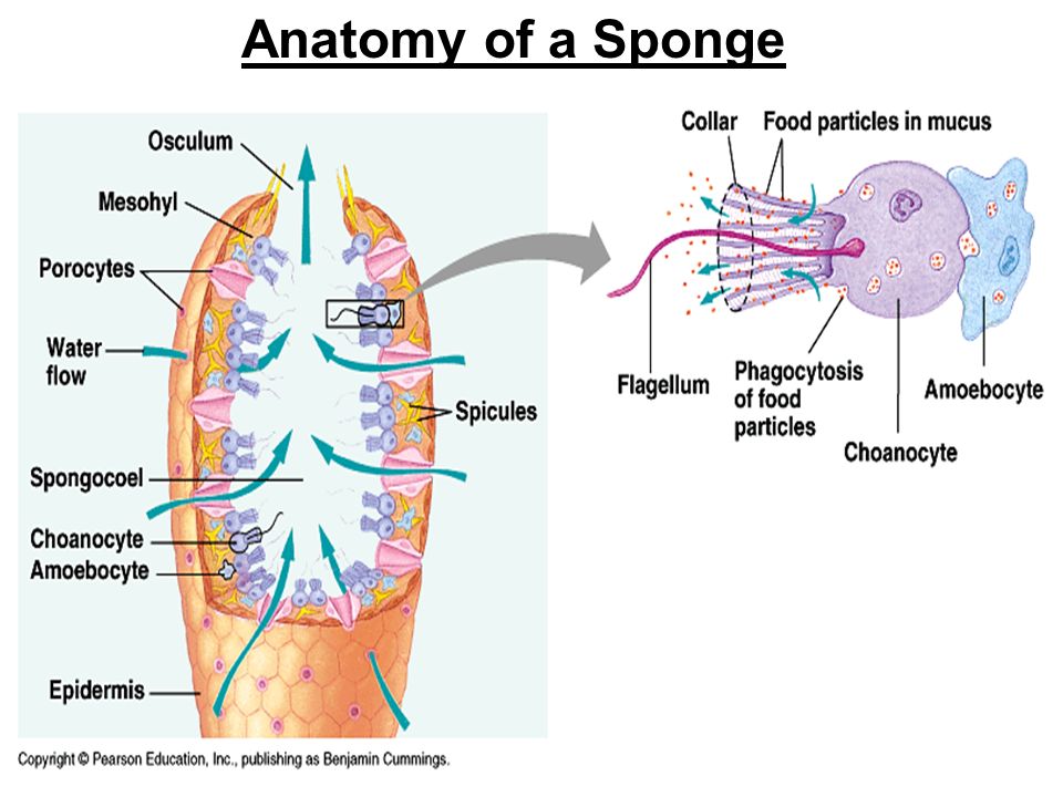
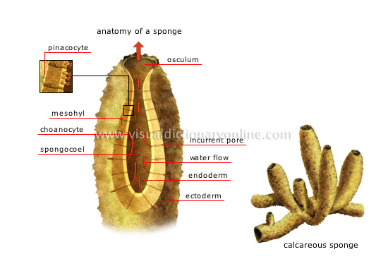
They are multicellular organisms that have bodies full of pores and channels allowing water to circulate through them consisting of jelly like mesohyl sandwiched between two thin layers of cells. Porifera is a phylum comprising of the multi cellular invertebrate animals called sponges.
 11 Best Phylum Porifera Images Sea Sponge Marine Biology
Sponges obtain nourishment and oxygen from this flowing water.
Sponge anatomy. Simple worksheet for labeling the parts of the sponge such as the osculum and choanocyte. These formations are made of a sponge like tissue containing trabeculae irregular blood filled spaces lined by endothelium and separated by connective tissue septa. Test your knowledge about sponge anatomy with this online quiz.
Simple sponges with choanocytes lining inner cavity radially symmetrical syconoid condition choanoderm and pinacoderm folding creates the shape. The branch of zoology that studies sponges is known as spongiology. A trivia quiz called sponge anatomy.
Wall of a sponge contains amoebocytes they are creeping mobile cells with a variety of functions including digestion and ability to differntiate into other cell types as needed. The term porifera literally means pore bearers. The flowing water also carries out waste products.
Cells embedded in the sponge tissue that ppick up nutrients from the incoming water and distribute nutrients throughout the sponge. Compressed polygonal cells called pinacocytes make up the pinacoderm the external sac layer. In the gel layer are either spicules supportive needles made of calcium carbonate or spongin fibers a flexible skeletal material made from protein.
The animals of this phylum have tiny pores in their body walls and this characteristic feature is the basis of the name of this phylum. The body of a sponge has two outer layers separated by an acellular having no cells gel layer called the mesohyl also called the mesenchyme. Choanocytes in small chambers called choanocyte chambers original much that open to an apopyle leading to the spongocoel.
The two corpora cavernosa lie along the penis shaft from the pubic bones to the head of the penis where they join. Sponges contain no organs or even tissue. Instead they consist of three cell sized layers.
The cells in the outer layers can move inward and change function.
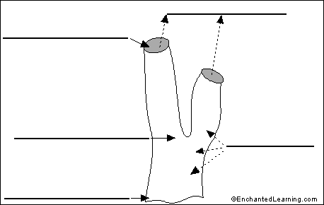
Label Sponge External Anatomy Enchantedlearning Com
 Phylum Porifera Sponge Anatomy Diagram Quizlet
 Papua New Guinea Png Milne Bay Province Coral Diversity
 Sponge Wikipedia
 Modern Sponge Anatomy A Schematic Cross Section Of
 C 1894 Sponge Anatomy Lithograph Original Antique Print Ocean Animal Sea Life Marine Fauna
 Sponge Wikipedia
 Ppt Sponge Powerpoint Presentation Free Download Id 1888638
 Sponge Anatomy Coloring And Information Sheet Anatomy
 Sponge Anatomy Science Biology Showme
 Sponge Gross Anatomy And Distinguishing Features Traits
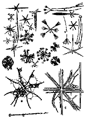
 Spicules Reproductive Biology Guws Medical

 Sponge Anatomy Chapter 24 Diagram Quizlet

 Anatomy Of Sponge Sea Sponge Anatomy Life Science
 Calcareous Sponges Reproductive Biology Guws Medical
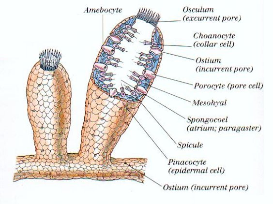
 Figure 1 From Metagenomic Approaches To Exploit The
 Sponge Anatomy Travel Mug By Bluespecsstudio
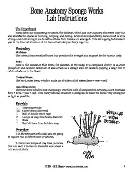 Bone Anatomy Sponge Works Human Body
 Sea Sponge Anatomy Zipper Pouch
 Classes Of Sponges And Sponge Anatomy Notes
 Review Of Animal Phylogeny Sponges Anatomy Of A Sponge

 Ppt Origins Of Multicellular Animals Powerpoint
 Sponge Sea Sponge Anatomy Biology
 Sponge Anatomy Coloring Color Anatomy
 Sponges General Anatomy
 Generalized Sponge Anatomy
Belum ada Komentar untuk "Sponge Anatomy"
Posting Komentar