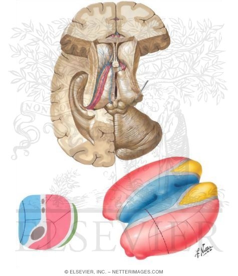
Anatomy Of The Thalamus
The thalamus translates neural impulses from various receptors to the cerebral cortex. Anatomy of the thalamus.
 Thalamus
In the rostral part of the thalamus the internal medullary lamina splits to form a partial capsule around the anterior nuclear group.
Anatomy of the thalamus. The thalamus has four surfaces medial lateral superior and inferior surface and it has two ends or poles anterior and posterior. In addition to the tracts mentioned above. Nuclei in a given pole or surface regulate specific functions or processing of sensory information and maintain particular connections with parts of the nervous and limbic system.
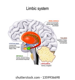
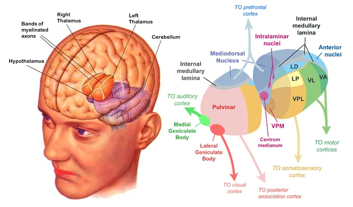
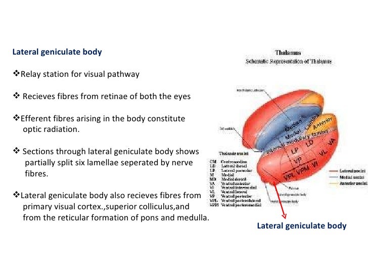
The posterior end of the thalamus is expanded to form the pulvinar. The external lamina covers the lateral surface and the internal lamina divides the nuclei into anterior medial and lateral groups. The thalamus is a limbic system structure and it connects areas of the cerebral cortex that are involved in sensory perception and movement with other parts of the brain and spinal cord that also have a role in sensation and movement.
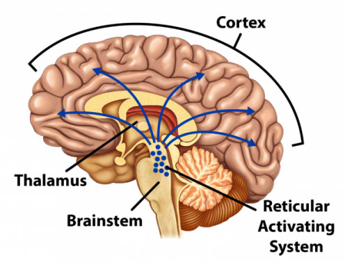
The anterior end of the thalamus is rounded and narrow which forms the posterior boundary. The thalamus separating it into medial and lateral nuclear masses. As a regulator of sensory information the thalamus also controls sleep and awake states of consciousness.
The lateral mass contains the lateral nuclear group and the ventral nuclear group. The medial mass consists of the medial nuclear group. Anatomy of the thalamus the thalamus has two ends the anterior and posterior poles and four surfaces.
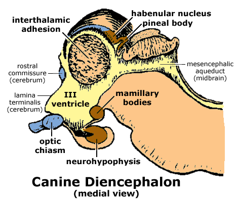
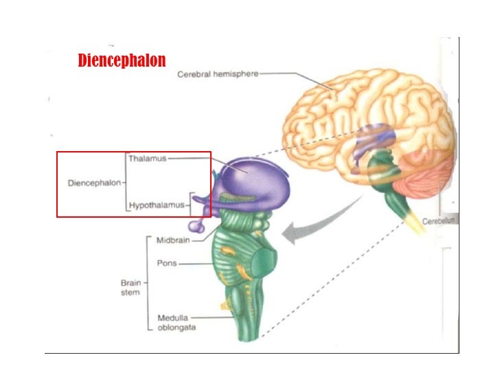
The thalamus is made up of two symmetrical structures formed from the diencephalon. The medial surface. The thalamus lies at the core of the diencephalon.
Medial lateral superior and inferior. Thalamus anatomy the thalamus a paired structure walnut sized shaped of grey matter found in the forebrain that is superior to the midbrain roughly the middle of the brain. Thalamus plural thalami either of a pair of large ovoid organs that form most of the lateral walls of the third ventricle of the brain.
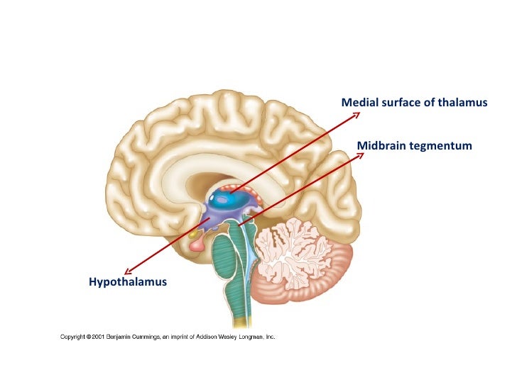
The inferior surface of the thalamus is continuous with the tegmentum of the midbrain. In addition to being divided into anterior. There are areas of white matter in the thalamus including the stratum zonale that covers the dorsal surface and the external and internal medullary laminae.
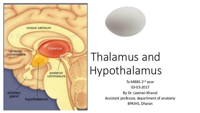
 Thalamus Hypothalamus
 What Is Thalamus And How It Looks Like
 The Thalamus And Hypothalamus Clinical Gate
 Thalamus Facts Position In Brain Summary Function
Antomy Of Thalamus And Hypothalamus
 Thalamus Brain Anatomy Medical Anatomy Psychiatry
 Thalamus Anatomy Location Structure Function Physiology

 Anatomy Of Thalamus
 What Is Thalamus And How It Looks Like
 Drawing Of The Brain Showing The Basal Ganglia Abd Thalamic Nuclei
 Thalamus Images Stock Photos Vectors Shutterstock

 Thalamus

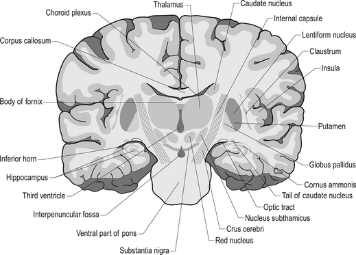
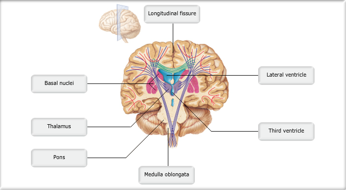
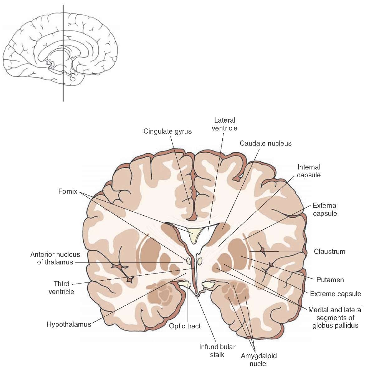
![]() Diagram Pictures Coronal Section Of The Brain At The
Diagram Pictures Coronal Section Of The Brain At The
 Anatomy Of The Brain Brain Anatomy And Disorders Of
 Thalamus Facts Position In Brain Summary Function
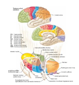
 Anatomy Of The Intralaminar And Medial Thalamic Nuclei And
 Anatomy Of Cerebral Cortex Thalamus Interconnexions J
 Print Anatomy And Physiology 1 Chapter 12 Flashcards Easy
 Median Section Of Human Brain Anatomical Structure
 Thalamus Hypothalamus Flashcards
 Brain Anatomy Stroke Emergency Care And Rehabilitation
 Thalamus Functions Of Thalamus Anatomy Clinical Significance
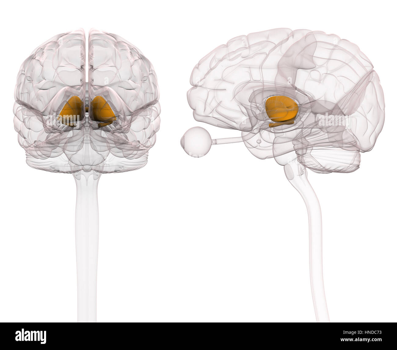 Thalamus Brain Anatomy Stock Photo 133675335 Alamy
![]() Thalamic Nuclei Connections Functions And Anatomy Kenhub
Thalamic Nuclei Connections Functions And Anatomy Kenhub
 Anatomical Illustrations Chelseacanlas
 Grey Matter Nuclei Non Cranial Nerve Thalamus
 Thalamus Definition Functions Location
 Anatomy Of Thalamus
 Anatomy Of Thalamus
Belum ada Komentar untuk "Anatomy Of The Thalamus"
Posting Komentar