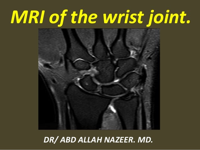
Wrist Mri Anatomy
Injuries such as anterior cruciate ligament meniscus and rotator cuff tears are all easily diagnosed when there is a firm understanding and knowledge of human anatomy. Effect on clinical diagnosis and patient care.
![]() Medical Imaging And Radiological Anatomy X Ray Ct Mri
Medical Imaging And Radiological Anatomy X Ray Ct Mri
With improved mri techniques the radiologist can increasingly visualize these ligaments.

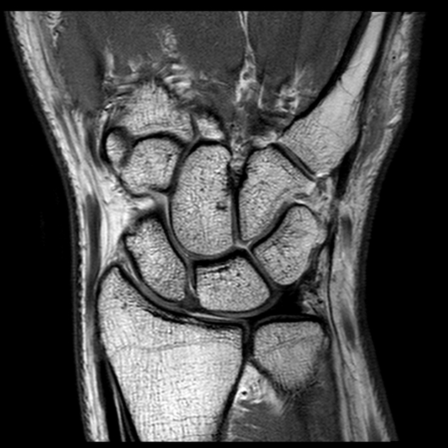
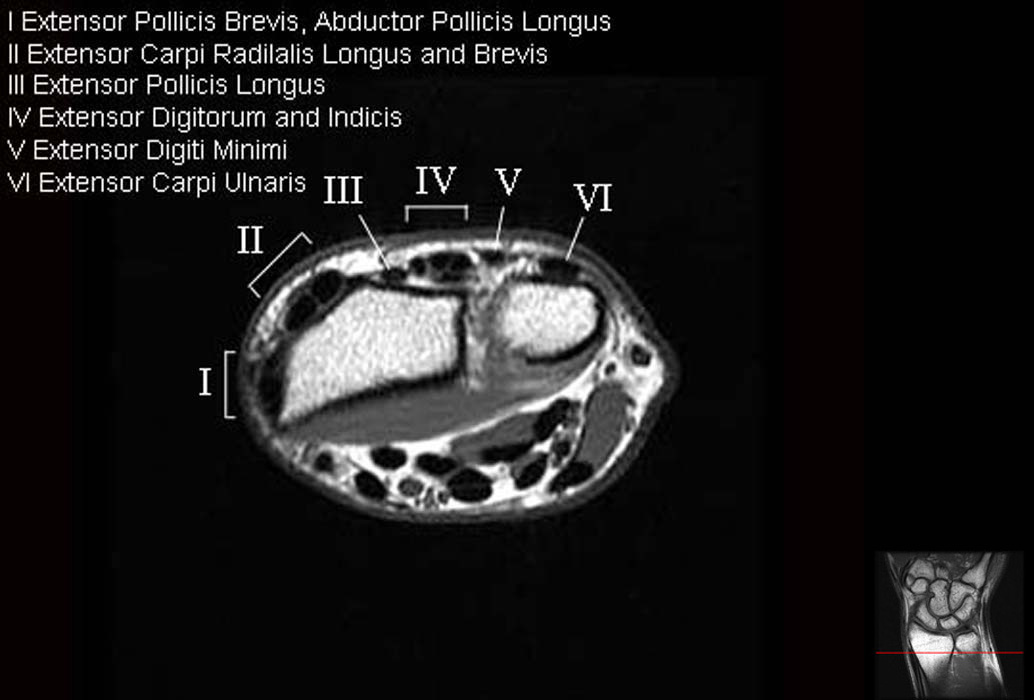
Wrist mri anatomy. 6 extensor pollicis longus tendon. Normal radiographic anatomy of the wrist. All the ligaments of the wrist visible in mri are shown on this anatomical module.
References hobby jl dixon ak bearcroft pw et al. 5 extensor digitorum indicis tt. About anatomy mri magnetic resonance imaging is particularly well suited for the medical evaluation of the musculoskeletal msk system including the knee shoulder ankle wrist and elbow.
A scaphoid fracture is the most common carpal fracture occurring more in active men. Use the mouse to scroll or the arrows. 1 flexor carpi ulnaris m t.
8 extensor carpi radialis brevis t. Unable to process the form. 4 extensor digiti minimi t.
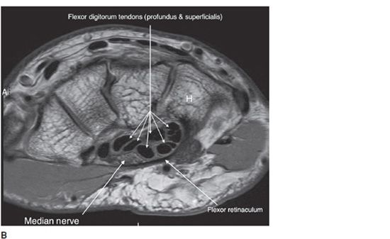
The anatomy mri appearance and clinical significance of the scapholunate ligament lunotriquetral ligament triangular fibrocartilage complex carpal metacarpal ligaments and volar and dorsal extrinsic ligaments are reviewed. Mr imaging of the wrist. The many tendons of the wrist are all captioned on this picture.
The distal radius and ulna articulate with the proximal row of the carpal bones consisting. Check for errors and try again. 9 extensor carpi radialis longus t.
3 extensor carpi ulnaris t. Use the mouse scroll wheel to move the images up and down alternatively use the tiny arrows on both side of the image to move the images. In recent years magnetic resonance imaging.
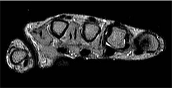
This mri wrist axial cross sectional anatomy tool is absolutely free to use. Mainly the muscles of the forearm and the palmar region muscles of the little finger. To sum up mri of the wrist is a relevant tool for diagnosis and clinical management of wrist pain including the evaluation of traumatic injuries and chronic syndromes.
 Accessory Muscles Of The Hand And Wrist Radsource
 Normal Wrist Mri Radiology Case Radiopaedia Org
 Mri Wrist Anatomy
 Wrist Anatomy Mri Wrist Axial Anatomy Free Cross
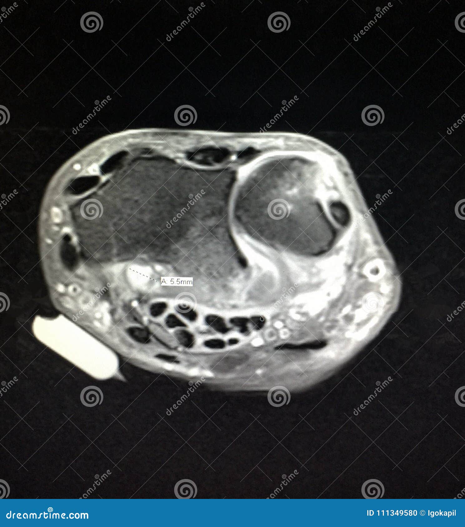 Radiological Mri Exam Wrist Anatomy Pathology Stock Photo
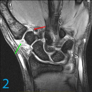
Mri Wrist Coronal Anatomy Wrist Tendon And Ligaments
![]() Medical Imaging And Radiological Anatomy X Ray Ct Mri
Medical Imaging And Radiological Anatomy X Ray Ct Mri
 Wrist Mri Radiology Key
 Presentation2 Pptx Wrist Joint
 Tfcc Tear Raleigh Hand Surgery Joseph J Schreiber Md
A Radiologist S Guide To Wrist Alignment
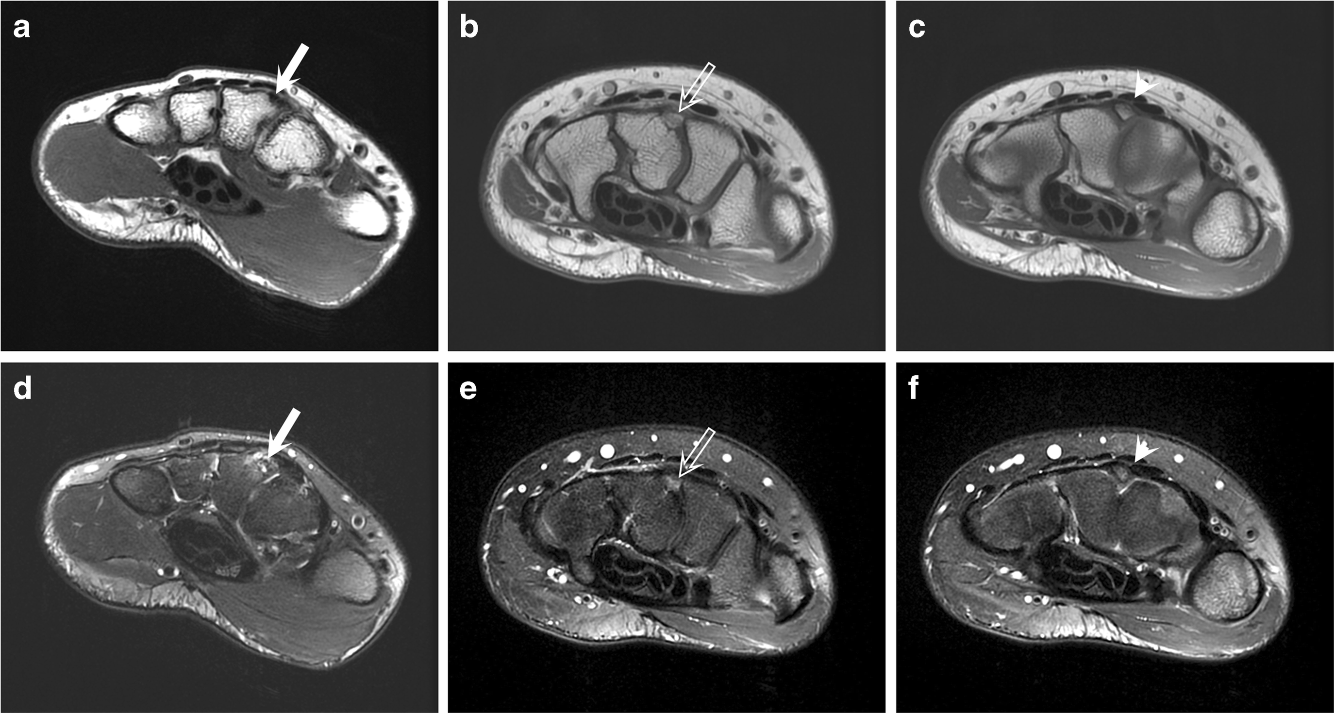
 Comparison Of Conventional Mri And Mr Arthrography In The
 Mri Of A Painful Carpal Boss Variations At The Extensor
 Anatomy Is King Or Queen In This Wrist Case
 Figure 11 From Imaging Of Radial Wrist Pain I Imaging
 Mri Anatomy Of Tfcc
 Mri Mastery Series Wrist Mri Online
 Aspetar Sports Medicine Journal Wrist Ligament Injuries In
 Wrist Mri
 Mri Technique Startradiology
 The Radiology Assistant Brain Anatomy
 Figure 4 From Ulnar Sided Wrist Pain Part I Anatomy And
 Wrist Mri Basic Musculoskeletal Imaging
 Mri Wrist Coronal Anatomy Wrist Tendon And Ligaments
 Wrist Anatomy Mri Wrist Axial Anatomy Free Cross
 Wrist Block Landmarks And Nerve Stimulator Technique Nysora
 Mri Wrist Coronal Anatomy Wrist Tendon And Ligaments
 Entrapment And Traumatic Neuropathies Of The Elbow And Hand


Belum ada Komentar untuk "Wrist Mri Anatomy"
Posting Komentar