Septum Anatomy
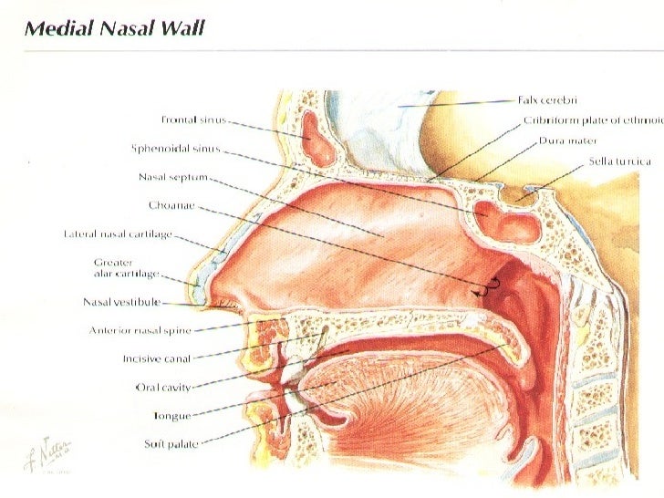
It consists of double layer. Cartilaginous and bony wall the nasal septum.
 Diagram Of The Nasal Septum
Called also septum lucidum.
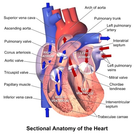
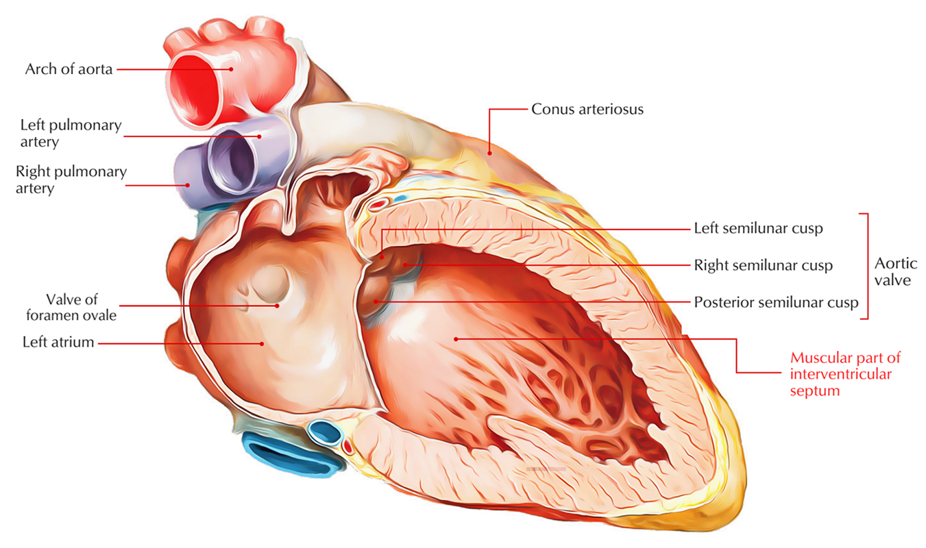
Septum anatomy. The floor of the nasal cavity is formed by the palate which also forms the roof of the oral cavity. The atria receive blood from various parts of the body and pass it into the ventricles. The apical two thirds of the ventricular septum is heavily trabeculated with a smooth walled basal one third 6.
The thin wall which separates the alveoli from each other in the lungs. Septum pellucidum or septum lucidum a thin structure separating two fluid pockets in the brain. It is formed of columella.
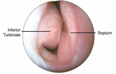
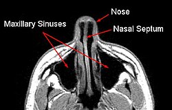
A thin wall called the septum divides the nose. Septum nasi separates the left and right nasal cavities. Anteriorly in an antero superior direction inferiorly rightward and inferolateral direction and apically toward the left ventricular apex.
See also congenital heart defect. Pellucid septum septum pellucidum the triangular double membrane separating the anterior horns of the lateral ventricles of the brain. The lower chambers the ventricles are separated by the interventricular septum.
Uterine septum a. Orbital septum a palpabral ligament in the upper and lower eyelids. Anteriorly the septal cartilage or quadrangular cartilage which approximates a quadrilateral shape.
Most of the sinuses drain into the nose through a small channel or drainage pathway that doctors call the middle meatus why do we have sinuses. It extends from the nares anteriorly to the choanae posteriorly and is covered by squamous epithelium. Nose anatomy nasal septum.
Roughly triangular in shape the septum extends in three planes. It consists of osteocartilaginous. A partition known as the interatrial septum.
The ventricles in turn pump blood to the lungs and to the remainder of the body. The nasal septum is composed of four structures. Each canal opens to the face by a nostril and into the pharynx by the choana.
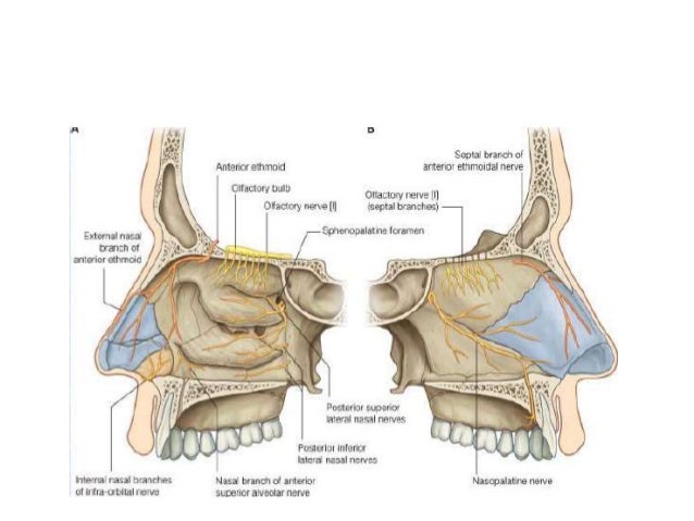
The above four arteries form a plexus in the anterioinferior portion of the nasal septum called the kiesselbochs plexus or little area. Human anatomy alveolar septum. The nasal septum latin.
Perpendicular plate of ethmoid bone. Septum primum a septum in the embryonic heart dividing the primitive atrium into right and left chambers. The vertical midline nasal septum is comprised primarily of a single nasal cartilage from the external nose and two bones.
 3 Blood Supply Of Nasal Septum From The Internet Euro
 Nose Bleed Management And Epistaxis Control Iowa Head And
 Rhinoplasty Anatomy
 Nasal Septum Septoplasty
 Right Ventricle Radiology Reference Article Radiopaedia Org
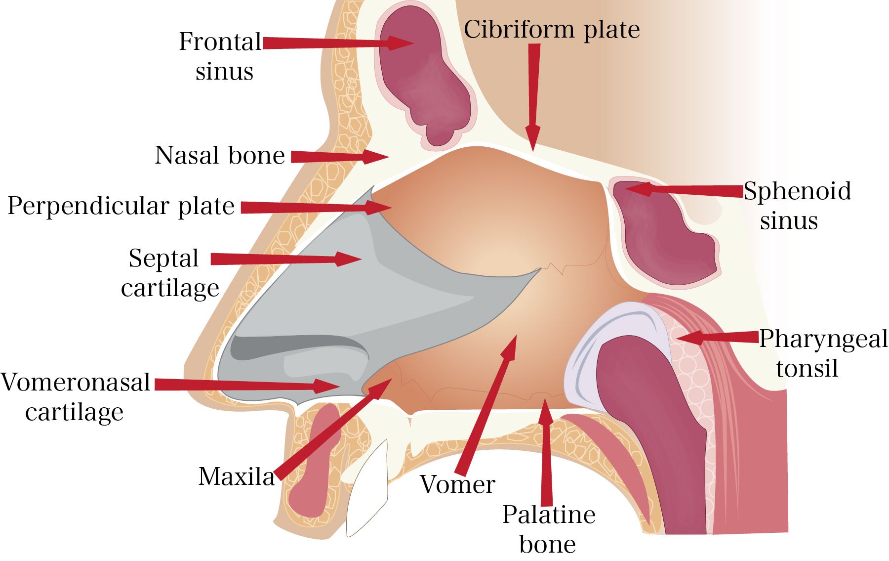 Anatomy Of The Nose Internal And External Nasal Structure
 Nasal Septal Perforation A Guide For Clinicians Consultant360
 Heart Septum Anatomy
 Nasal Airway Obstruction Ent Stryker
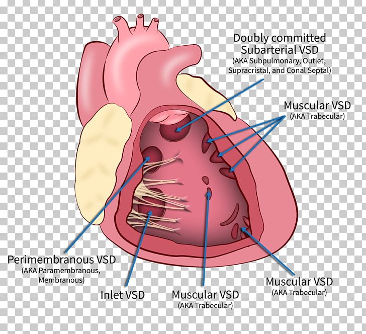
Heart Ventricular Septal Defect Interventricular Septum
 Nasal Obstruction Septoplasty Ent Of Georgia South
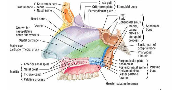
 Bones Of The Nasal Cavity Skull Anatomy Nasal Cavity
 Maldeveloped Septum Pellucidum Associated With Schizophrenia
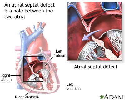 Atrial Septal Defect Asd Medlineplus Medical Encyclopedia
 Mercy Angiography Conditions Diseases Interventional
 Septoplasty New Jersey Deviated Septum Repair Princeton
 The True Interatrial Septum Near The Tricuspid Annulus Open I
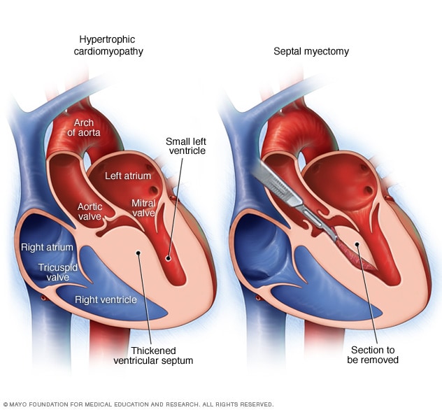 Septal Myectomy Mayo Clinic
 Nasal Physiology Overview Anatomy Of The Nose Nasal Airflow
 Anatomy Of The Left Ventricle Dr Sean Lal
 Interventricular Septum Earth S Lab
 Anatomy Nasal Septum And Septoplasty Pakistan
 Cardiac Interventions Today Transseptal Puncture A Step
 Nasal Septum Wikipedia
 Treatment Of Hematoma Of The Nasal Septum Nejm


Belum ada Komentar untuk "Septum Anatomy"
Posting Komentar