Retroperitoneal Anatomy
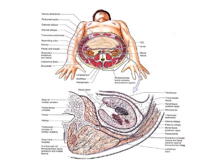
Topographic retroperitoneal anatomy the posterior abdominal wall is the posterior boundary of the abdominal cavity which is the continuous part of posterior thoracic wall from the level of diaphragm cranially and posterior pelvic wall caudally. Surgical exposureanatomy of the lateral lumbar spine and plexus.
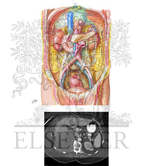
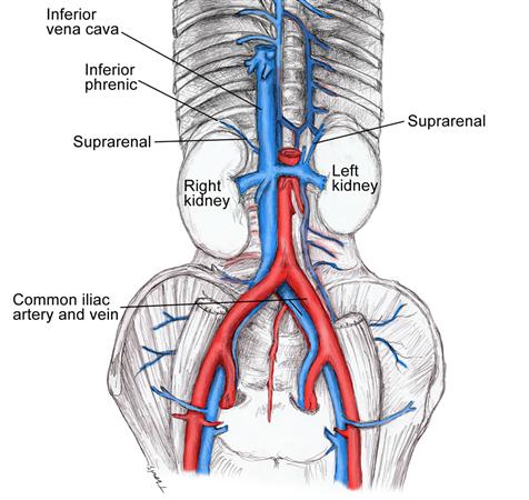
 Abdominal Retroperitoneal Anatomy Medical Illustration
It has become essential that radiologists thoroughly understand.

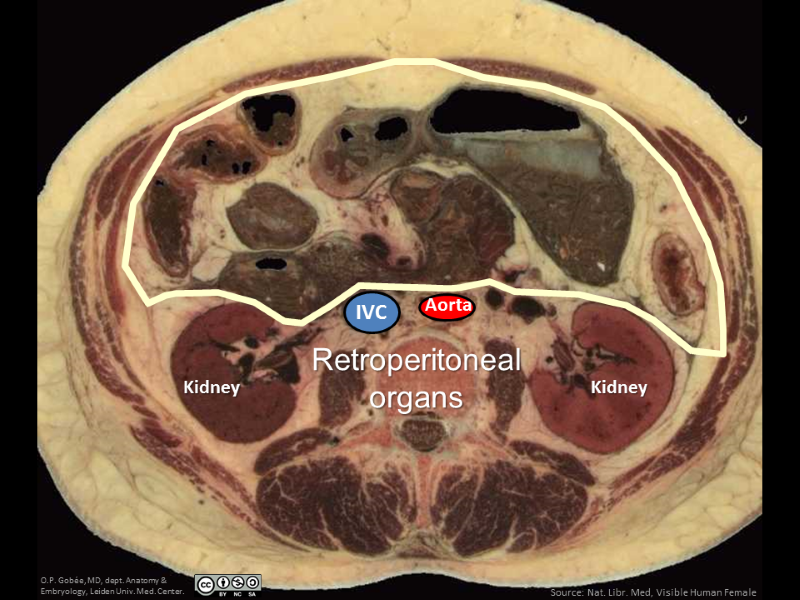
Retroperitoneal anatomy. Peritoneal and retroperitoneal anatomy and its relevance for cross sectional imaging learning objectives. Retroperitoneal structures include the rest of the duodenum the ascending colon the descending colon the middle third of the rectum and the remainder of the pancreas. Kidneys adrenals and retroperitoneum.
Secondarily retroperitoneal meaning the structures initially were suspended in mesentery and later migrated behind the peritoneum during development the duodenum except for the proximal first segment which is intraperitoneal. The retroperitoneal space is bounded by the posterior parietal peritoneum anteriorly. It extends from the 12th thoracic vertebra and 12th rib above to the sacrum and iliac crest below.
Retroperitoneal space is the anatomical space in the abdominal cavity behind the parietal peritoneum. The retroperitoneal space is mesodermally derived and is. Discuss the importance of identifying peritoneal anatomy in assessing extent.
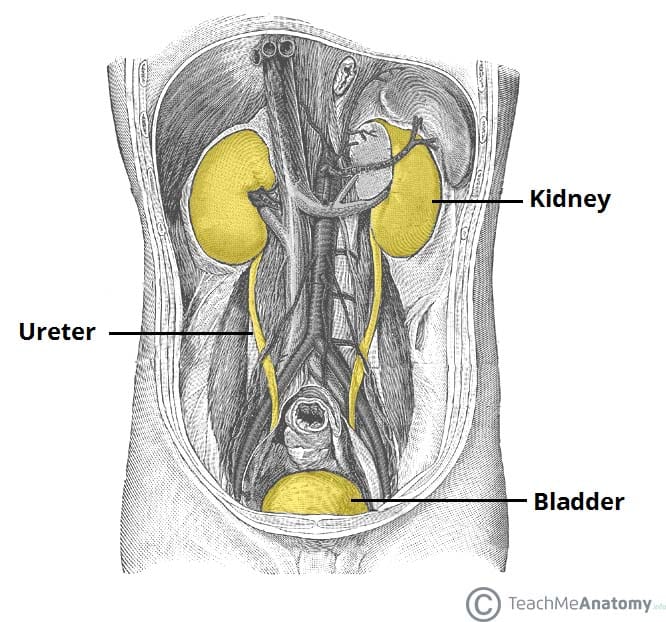
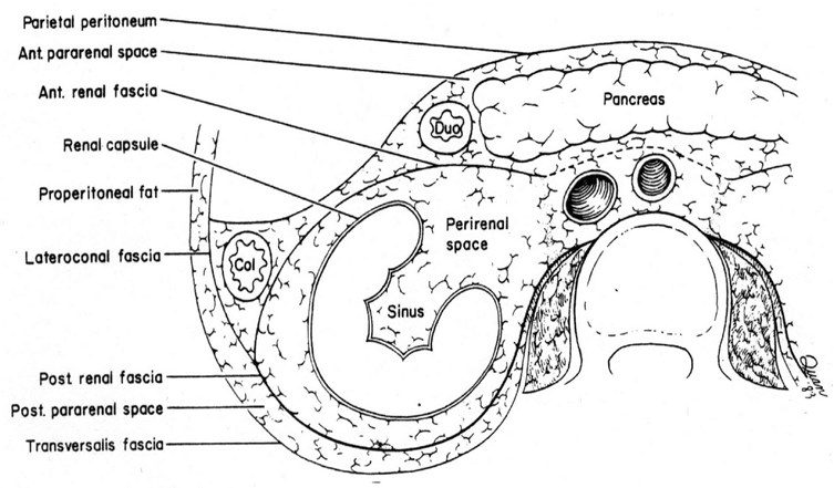
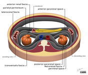
It is divided into three spaces by the perirenal fascia and is best visualized using ct or mri. Ascending and descending portions of the colon but not the transverse. Other organs located in the retroperitoneal space are the kidneys adrenal glands proximal ureters and renal vessels.
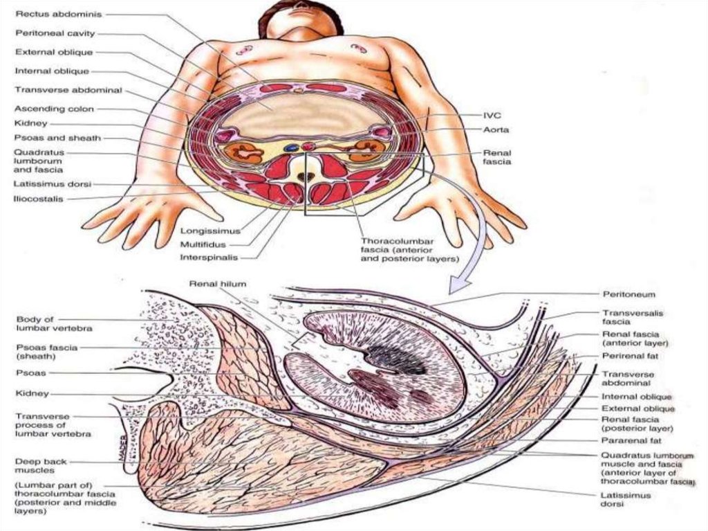
A useful mnemonic to remember which organs are retroperitoneal is. The retroperitoneum is the part of the abdominal cavity that lies between the posterior parietal peritoneum and anterior to the transversalis fascia. The floor of the space that is the posterior wall of the abdomen in this region is formed by.
Retroperitoneal organs mnemonic dr yuranga weerakkody and dr henry knipe et al.
 Normal Retroperitoneal Anatomy Medivisuals Medical
 Illustration Displaying Retroperitoneal Anatomy In Axial
 Reproteritoneum Anatomy And Pathology
 Search Retroperitoneal
 Extraperitoneal Retroperitoneal Subperitoneal
 Turkish Journal Of Obstetrics And Gynecology
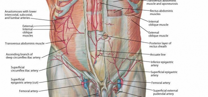
 Abdominal And Retroperitoneal Anatomy Medical Illustration
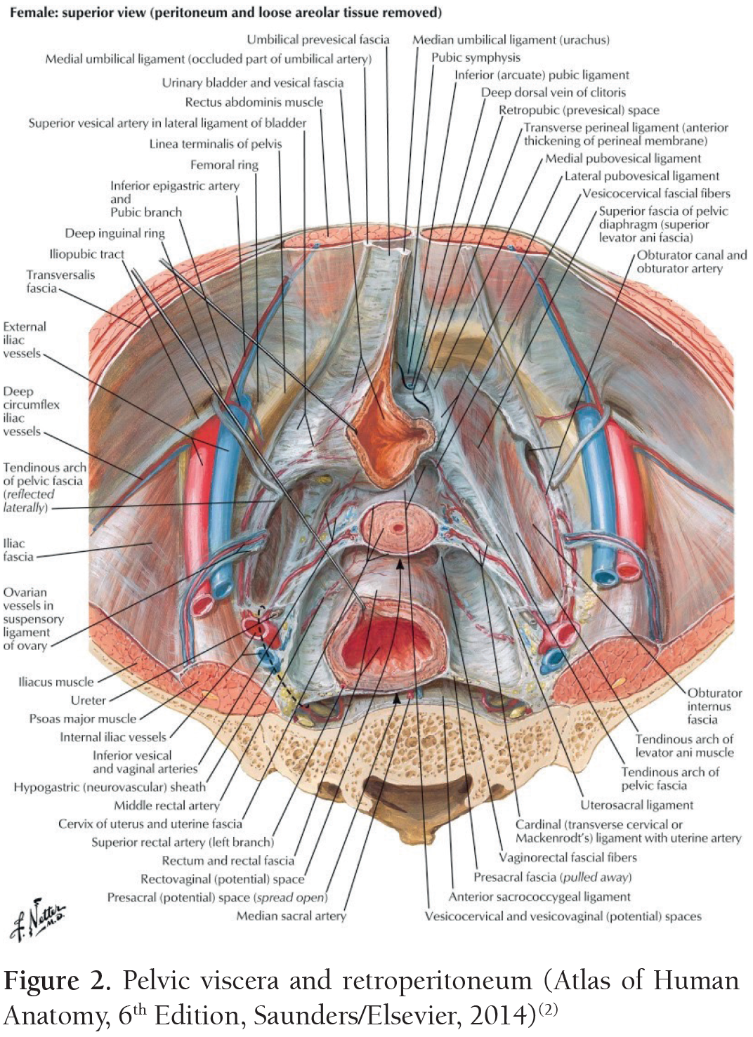
 Going Through Netter S To Teach Medical Students Pelvic Anatomy
 The Kidneys Position Structure Vasculature
 Retroperitoneal Anatomy And Ct Scan Of Large Tumor
 The Retroperitoneal Space Retroperitoneum Is The
 Abdominal Wall Omentum Mesentery And Retroperitoneum
 Osta Of Lumbar Retroperitoneal Space Online Presentation
 The Retroperitoneal Spaces Mh
 Retroperitoneal Anterolateral Approach To The Lumbar Spine
 Retroperitoneal Anatomy And Ct Scan Of Large Tumor
 Retroperitoneum Radiology Reference Article Radiopaedia Org
 Figure 6 From Understanding Retroperitoneal Anatomy For
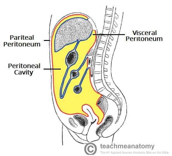 The Peritoneum Visceral Parietal Teachmeanatomy
 Internal Organs Atlas Of Anatomy
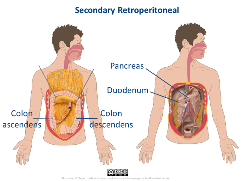 Secondary Retroperitoneal Anatomytool
![]() Peritoneum And Peritoneal Cavity Anatomy And Function Kenhub
Peritoneum And Peritoneal Cavity Anatomy And Function Kenhub
 Retroperitoneal Anatomy
Intra And Retroperitoneal Anatomy Landmarks And Pearls Of
 Radioanatomy Of The Retroperitoneal Space Sciencedirect
 Amicus Illustration Of Amicus Anatomy Abdominal Peritoneum
Retroperitoneal Structures Gastrointestinal Medbullets
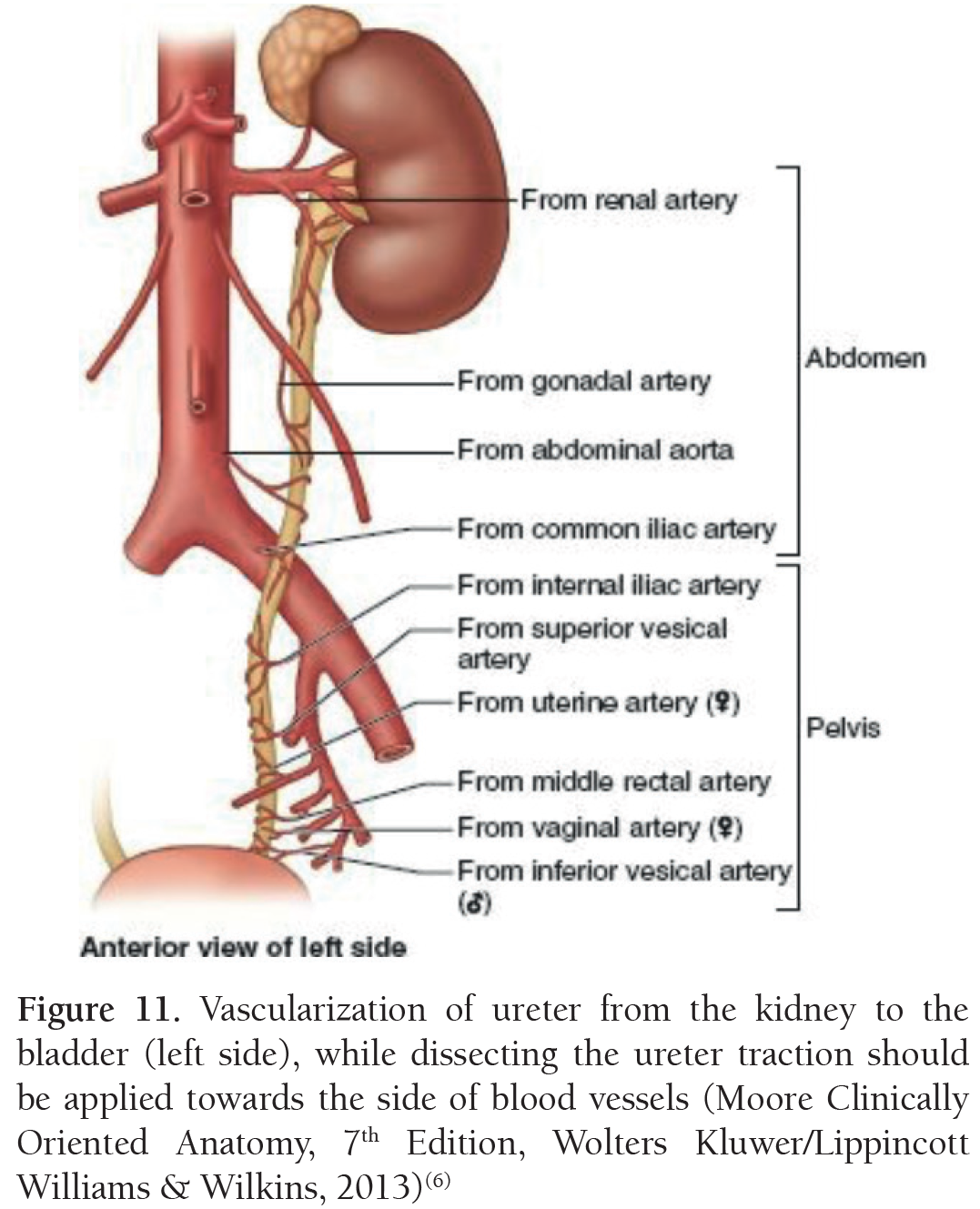 Turkish Journal Of Obstetrics And Gynecology
 Pin By Stelios Daskalogiannis On Peritoneum Retroperitoneum
 Clinically Oriented Anatomy Of Retroperitoneum Kidneys And
 What Urinary System Structure Is Located Retroperitoneal
 Peritoneal Cavity Part 4 Intraperitoneal And Retroperitoneal Organs Anatomy Tutorial
 Surgical Anatomy Of The Retroperitoneum Adrenals Kidneys


Belum ada Komentar untuk "Retroperitoneal Anatomy"
Posting Komentar