Cochlea Anatomy
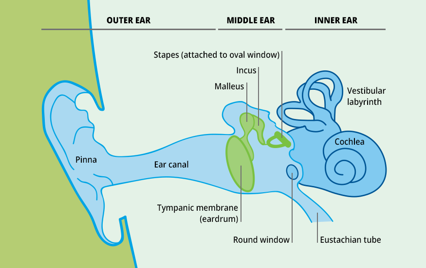
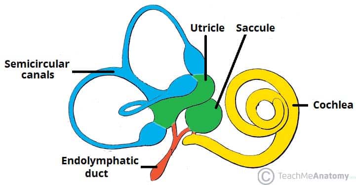
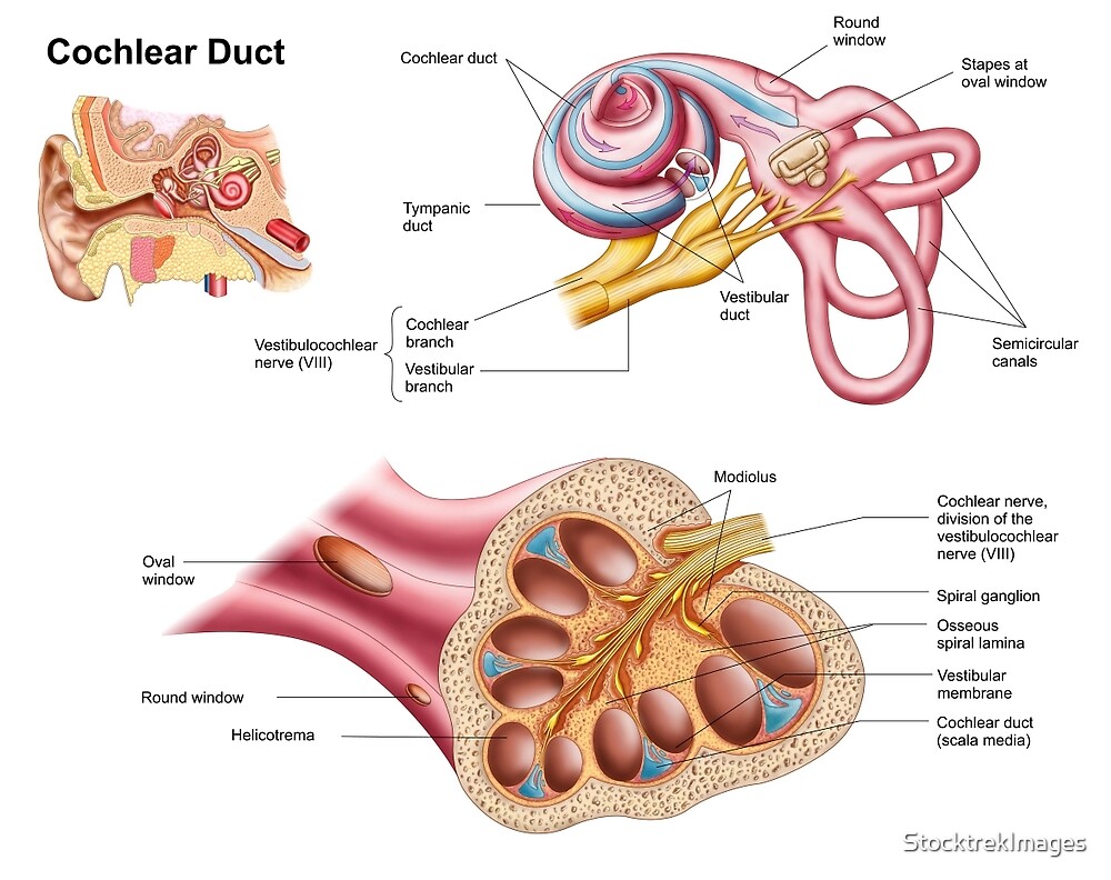
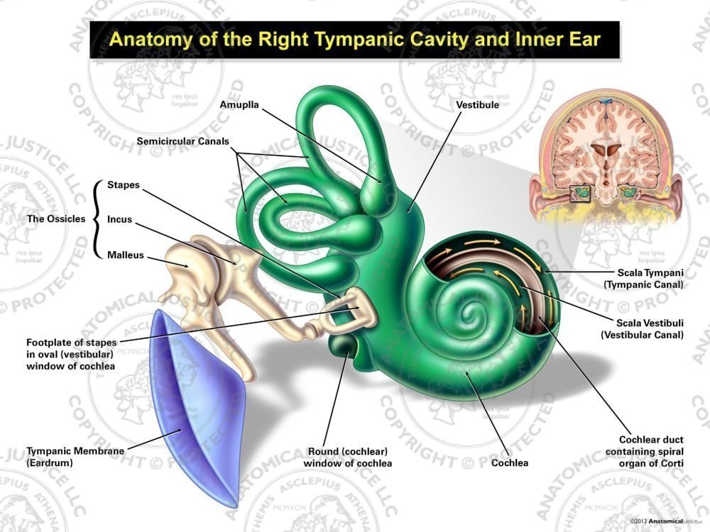
Cochlea anatomy the cochlea created by three basic chambers resembles a snail and wraps itself two and a half times around the central bone of the core. The cochlea is the main structure of the human auditory system.
Auditory And Vestibular Systems Structure And Operation Of
The basilar membrane a main structural element that separates.
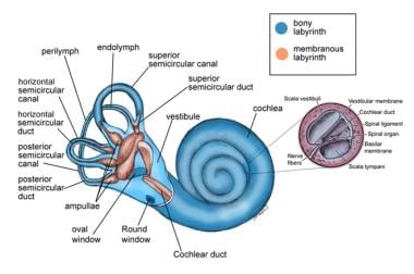
Cochlea anatomy. 0 0000 a shoutout is a way of letting people know of a game you want them to play. It contains the cochlear duct part of the membranous labyrinth which senses hearing. The cochlear structures include.
Physiology of hearing in senses. Choose from 461 different sets of cochlea anatomy flashcards on quizlet. Reissners membrane which separates the vestibular duct from the cochlear duct.
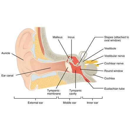
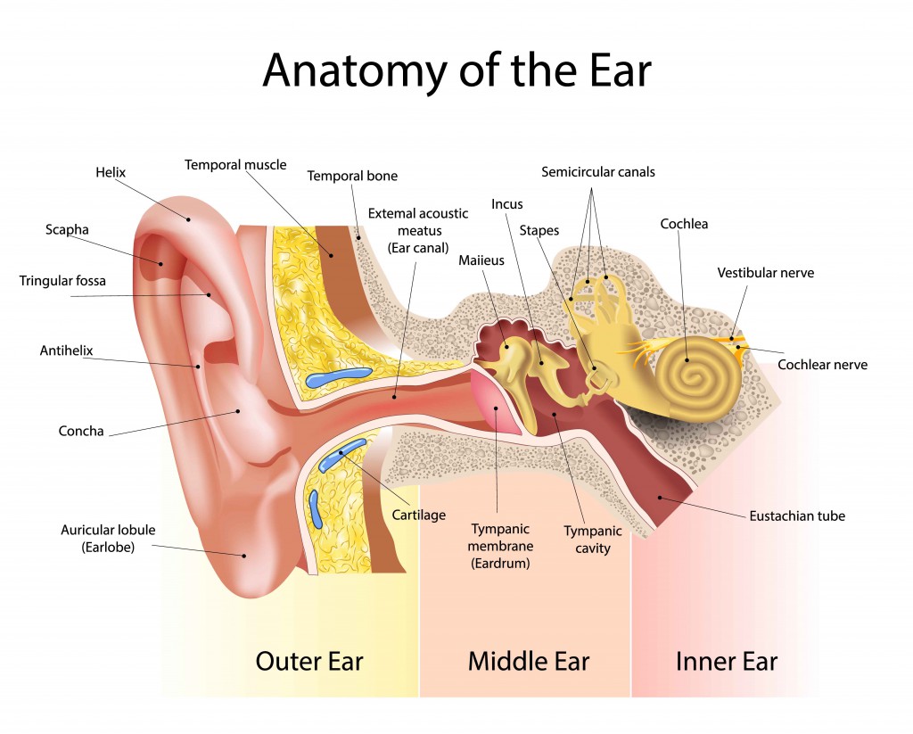
Gross anatomy the cochlea is a shell shaped spir. It is not actually an organ itself but a bony structure in the inner ear that contains the auditory organ. Sound waves travel into the outer ear canal vibrating the structures of the air filled middle air which transmit the waves to the fluid of the inner ear by the stapes bone hitting a membrane called the oval window.
The cochlea has a base screw head and an apex point. It was created using 3ds max premier pro after effects garage band and sound bo. The helicotrema the location where the tympanic duct and the vestibular duct merge.
Cochlea structure the lower chamber scala tympani can be traced from the apex to the cochlear window. The cochlea is a complex coiled structure. Learn cochlea anatomy with free interactive flashcards.
Just pick an audience or yourself and itll end up in their incoming play queue. Cochleae is part of the inner ear osseous labyrinth found in the petrous temporal bone. Mechanical senses inner ear which contains the cochlea.
The bony spiral lamina is represented by the threads of the screw. Three scalae or chambers. Cochlear anatomy the cochlea has a bony core called the modiolus.
This animation provides a general overview of the anatomy of the cochlea. The cochlea houses the cochlea duct of the membranous labyrinth the auditory part of the inner ear. At the vestibular window the first chamber scala vestibule reaches to the termination of the cochlea.
The modiolus defines the medial direction in the cochlea the outside wall is lateral. Transmission of sound waves in the cochlea the mechanical vibrations of the stapes footplate at the oval window creates pressure waves in. It twists upon itself around a central portion of bone called the modiolus producing a cone shape which points in an anterolateral direction.
Cochlear Anatomy Function And Pathology I
 Minimally Invasive Cochlear Implant Surgery Background
 Cochlea Radiology Reference Article Radiopaedia Org
 Anatomy Of The Organ Of Corti Part Of The Cochlea Of The Inner Ear Canvas Print
 Anatomy Of The Auditory System Ento Key
 The Cochlea Acland S Video Atlas Of Human Anatomy
 The Inner Ear Bony Labyrinth Membranous Labryinth
 Anatomy Of The Cochlear Duct In The Human Ear By
 Anatomy Of The Cochlea Cartoon Illustration Of The Cochlea
 Anatomy Of The Right Tympanic Cavity And Inner Ear

 Anatomy Hearing Part 2 Functions Of Cochlea Organ Of Corti
 Inner Ear Anatomy Physiology 2201 With Aldridge At
 Anatomy Of The Ear Inner Ear Middle Ear Outer Ear
 Amazon Com Cochlea Of Inner Ear Watercolor Art Print
 Inner Ear Discovery Helps Explain How Sound Waves Become
 Ear Anatomy Inner Ear Cochlea Histology Vestibular System Structure Cochlea Histology Audiology Medical Office Decor
 8 2 2 Anatomy Of Ear Inner Ear Diagram Quizlet
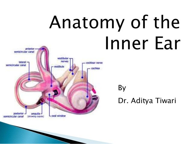 Anatomy Of Inner Ear By Dr Aditya Tiwari
 Cochlea Diagram Ear Anatomy Ear Function Eye Anatomy
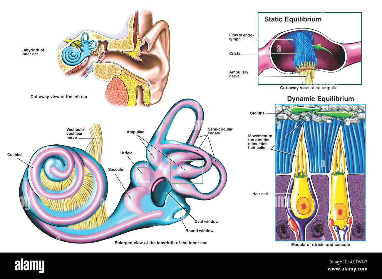 Anatomy Of The Inner Ear Stock Photo 7710486 Alamy
 Organ Of Corti Model With Representation In Cochlea 3b Smart Anatomy
 Anatomy Of The Cochlear Duct In The Human Ear By Stocktrek Images Art Print Poster
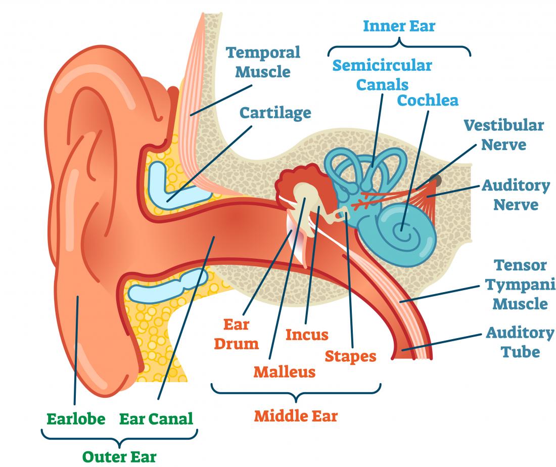 Ears And Hearing How Do They Work



Belum ada Komentar untuk "Cochlea Anatomy"
Posting Komentar