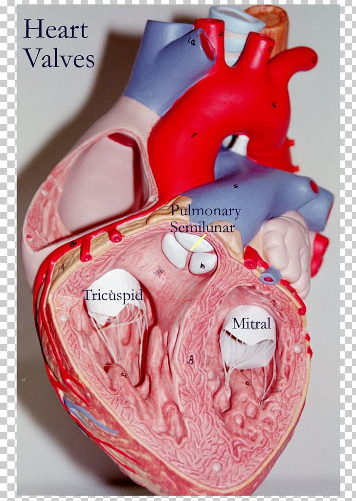
Heart Valves Anatomy
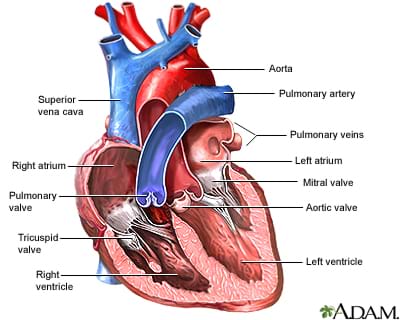
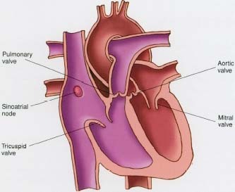
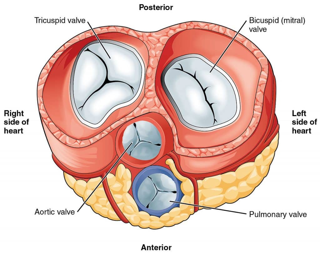
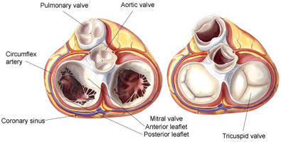
The pulmonary valve and aortic valve. Blood passes through a valve before leaving each chamber of the heart.
 Novel Technique Reduces Obstruction Risk In Heart Valve
They are located between the atria and corresponding ventricle.
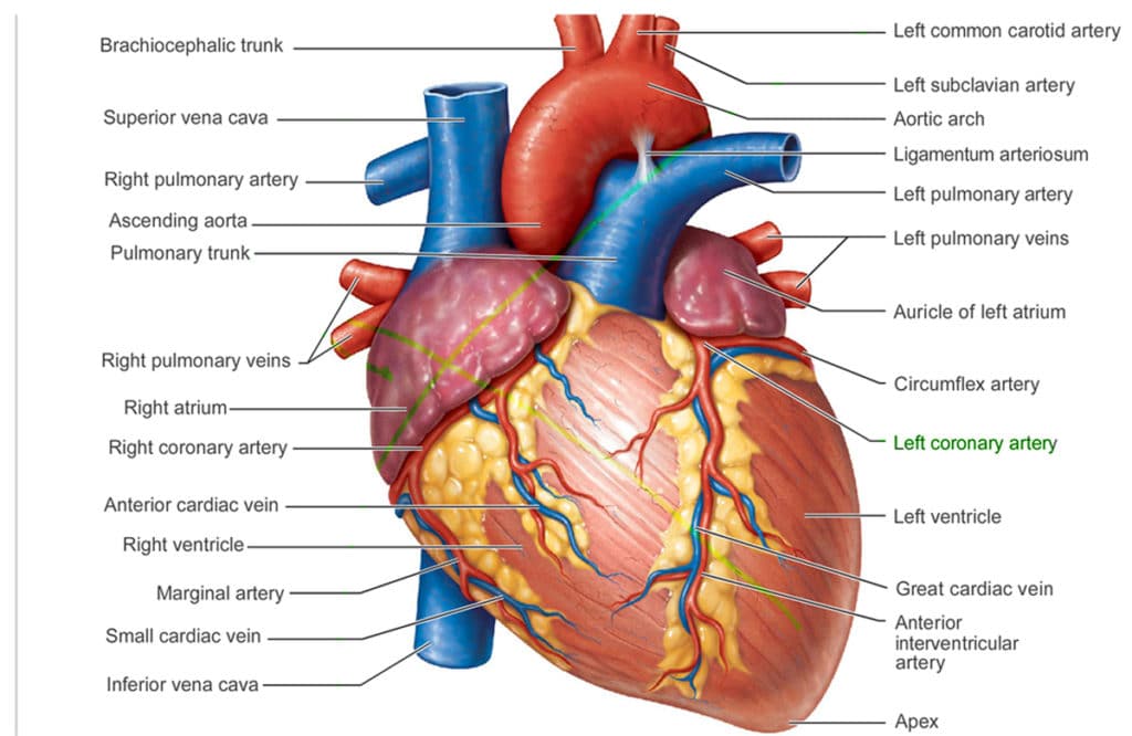
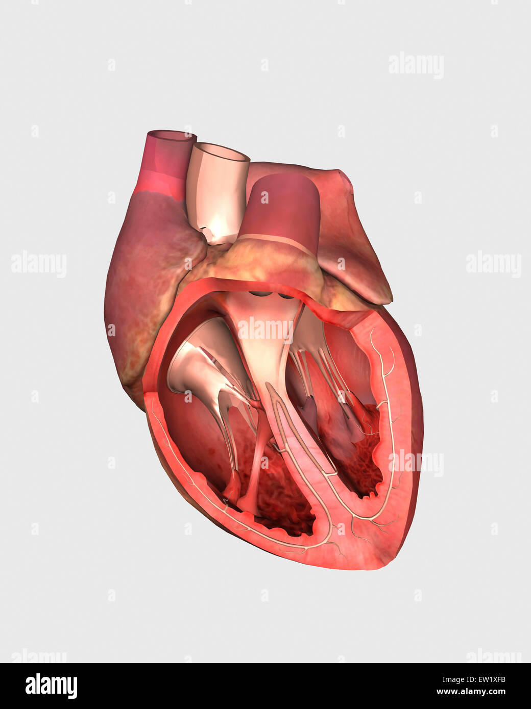
Heart valves anatomy. This article aims to explore the embryology and gross anatomy of the heart valves. The heart valves are uniquely designed gates that promote the unidirectional flow of blood through the heart. They are located between the.
The valve between the left atrium and the left ventricle is called the mitral valve. This heart valve is located between the right atrium and the right ventricle. When closed it allows oxygen depleted blood returning to.
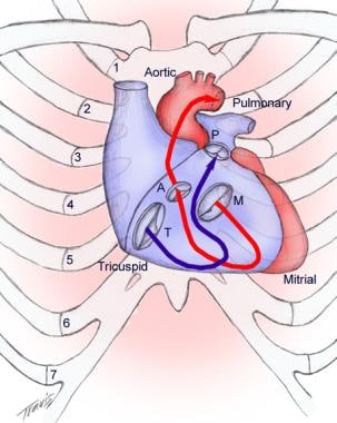
The tricuspid valve and mitral bicuspid valve. The four valves of the heart. Additional discussion about the heart sounds and their relationship to the status of the valves open or closed disorders that affect the valves valvulopathies or valvular heart disease and clinical examination of the.
This heart valve is located between the left atrium and left ventricle. They are composed mostly of fibrous connective tissue that extends from the heart walls. The valves prevent the backward flow of blood.
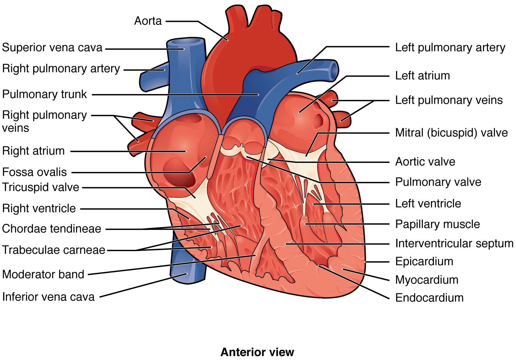
There are four valves of the heart which are divided into two categories. A valve may consist of a sphincter muscle or two or three membranous flaps or folds. The heart has 4 chambers 2 upper chambers atria and 2 lower chambers ventricles.
When closed it allows the. Introduction to the anatomy of the heart valves. The right atrioventricular valve or av valve controls blood flow from the right atrium to the right ventricle.
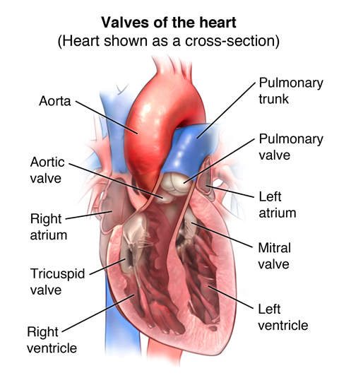
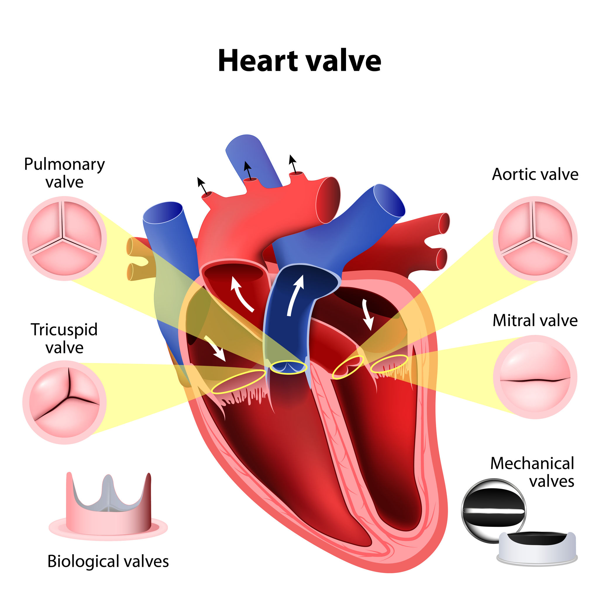
Semilunar valves control blood flow out of your heart. They are attached to special muscular appendages that help to keep them stable. Valve in anatomy any of various membranous structures especially in the heart veins and lymph ducts that function to close temporarily a passage or orifice permitting movement of a fluid in one direction only.
Anatomy and function of the heart valves what are heart valves. Atrioventricular av valves tricuspid valve. Blood flows out of the right ventricle to the lungs through the pulmonary valve.
The external surfaces of the valves are covered by endocardium. The valve between the right atrium and the right ventricle is called the tricuspid valve.
 Roles Of Your Four Heart Valves American Heart Association
 Heart Valves Yourheartvalve
 Heart Valves Function And Anatomy How The Heart Valves Work
 Heart Valve Anatomy
 What Do I Need To Know About Heart Valves Lesson
 Heart Anatomy Guide For Patient Consultation New Heart Valve
 Anatomy Of The Heart Heart Valves Function Purpose And How
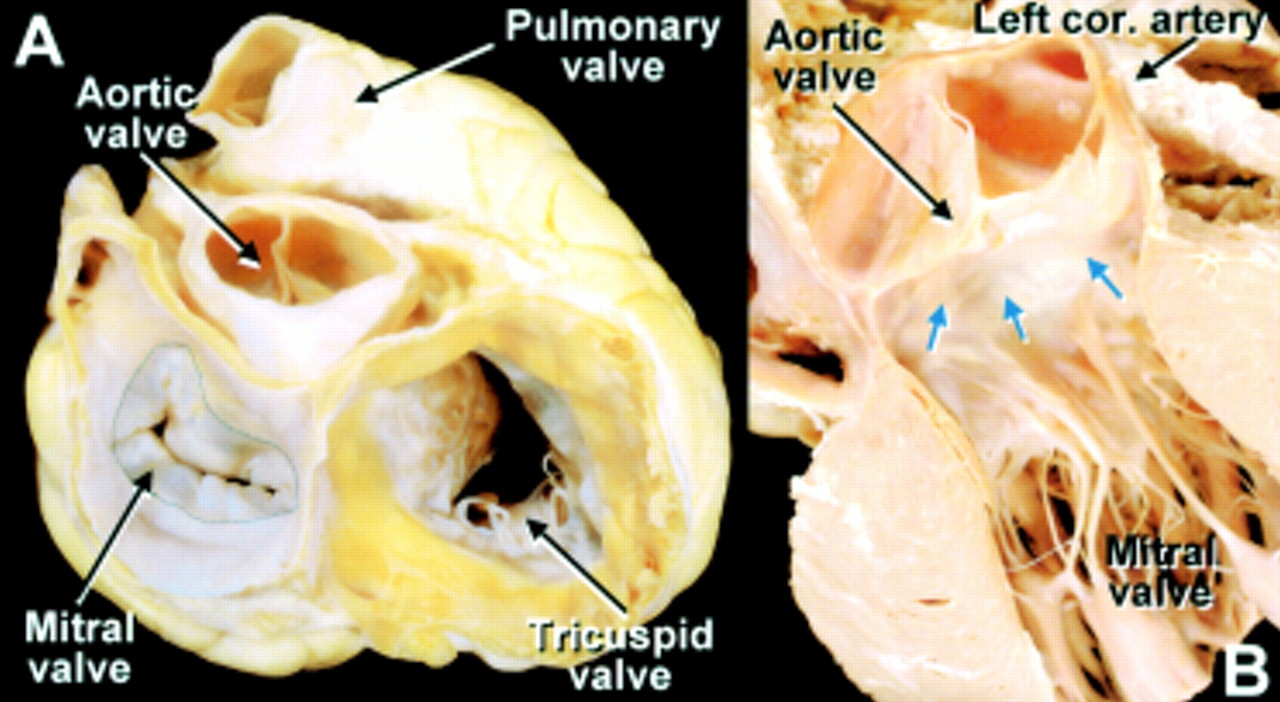
Schematics Of Heart Valve Anatomy A The Arrangement Of
 The Heart
 Heart Valves Anatomy
 Heart Valve Anatomy Tricuspid Valve Png Clipart Anatomy
 Heart Valves Anatomy Defects Disease Problems

 19 1 Heart Anatomy Anatomy And Physiology
 Anatomy Of The Heart Blood Flow Through The Heart And The
 Heart Valve Anatomy
 Anatomy Of The Mitral Valve Heart
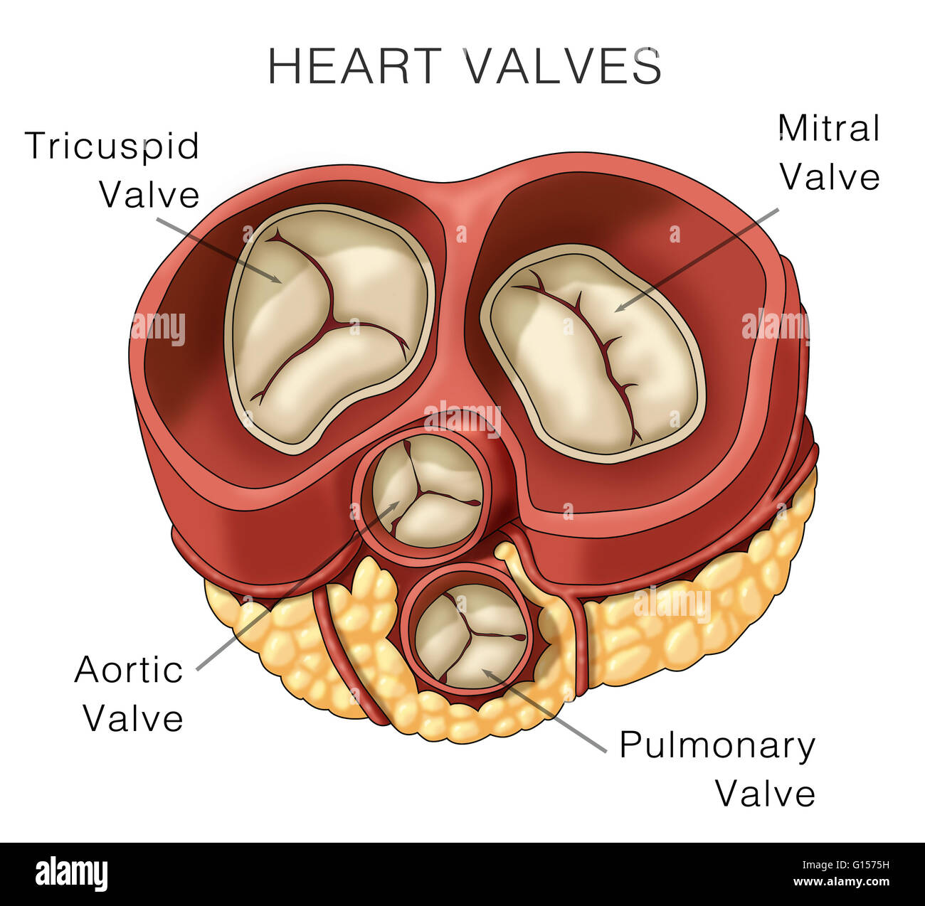 Illustration Of Heart Valves The Image Shown Includes The
![]() Heart Valves Anatomy Tricuspid Aortic Mitral Pulmonary Kenhub
Heart Valves Anatomy Tricuspid Aortic Mitral Pulmonary Kenhub
 Valves Of The Heart Preview Human Anatomy Kenhub
 Aortic Valve Anatomy The Aortic And Pulmonary Valves Are
 Tricuspid Valve Anatomy Overview Gross Anatomy
 A Beginner S Guide To Heart Anatomy Heart Surgery Information
Heart Valve Wikipedia
 Heart Valves Showing Pulmonary Valve Mitral Valve And
 Pin By India Cardiac Surgery Site On Low Cost Heart
 Acquired Heart Valve Disease Symptoms
 Heart Valve Wikipedia
 The Heart Valves Tricuspid Aortic Mitral Pulmonary
 An Overview Of Heart Valve Disease
Belum ada Komentar untuk "Heart Valves Anatomy"
Posting Komentar