Coxal Bone Anatomy
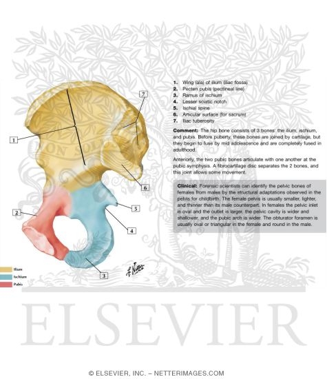
Anatomy of the hip. Prior to puberty the t riradiate cartilage separates these parts and fusion only begins at the age of 15 17.
 The Pelvis Human Anatomy And Physiology Lab Bsb 141
Look it up now.
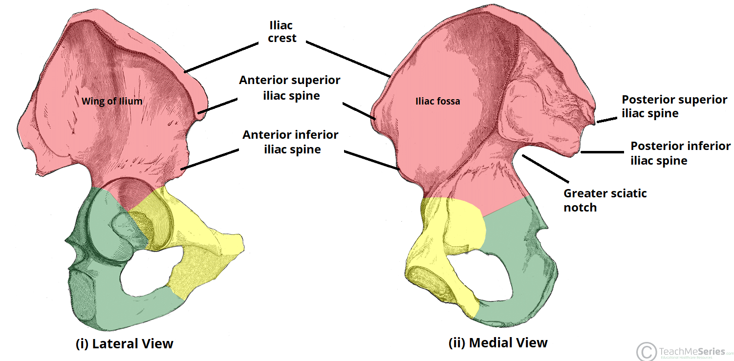
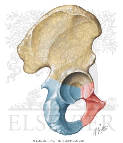
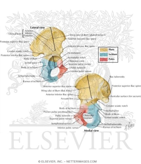
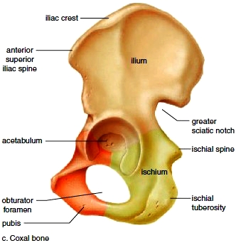
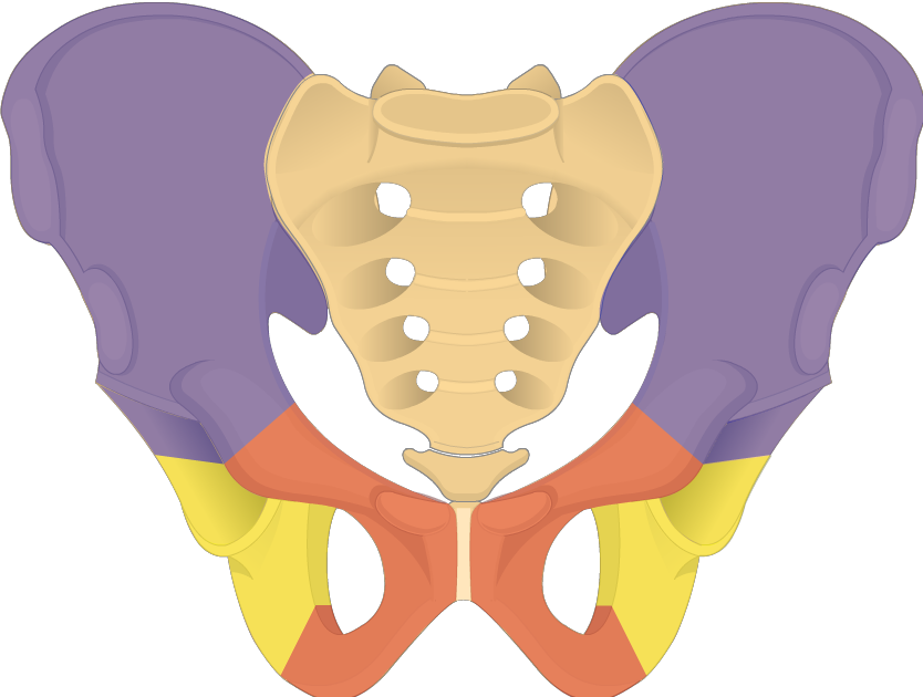
Coxal bone anatomy. If you think of the hip joint in layers the deepest layer is bone then ligaments of the joint capsule and the tendons and muscles are on top. The ilium ischium and the pubis. In some vertebrates including humans before puberty it is composed of three parts.
It meets its fellow on the opposite side in the middle line in front and together they form the sides and anterior wall of the pelvic cavity. The hip bone is comprised of the three parts. Just pick an audience or yourself and itll end up in their incoming play queue.
In adults three of the bones of the pelvis have fused into the hip bone or acetabulum which forms part of the hip region. The hip joint is a ball and socket synovial joint formed between the os coxa hip bone and the femur. Send a shoutout about this game.
Anatomy made simple by dr. Nerves and vessels supply the muscles and bones of the hip. The hip bone os coxae innominate bone pelvic bone or coxal bone is a large irregular bone constricted in the center and expanded above and below.
Coxae is the joint between the femur and acetabulum of the pelvis and its primary function is to support the weight of the body in both static eg. Together the ilium pubis and ischium form a cup shaped socket known as the acetabulum literal meaning in latin is vinegar cup. A shoutout is a way of letting people know of a game you want them to play.
Hip bone part 1 duration. Standing and dynamic eg. Coxal pelvis ap1 rita thrasher.
The hip joint scientifically referred to as the acetabulofemoral joint art. It bears our bodys weight and the force of the strong muscles of the hip and leg. Unsubscribe from rita thrasher.
Yet the hip joint is also one of our most flexible joints and allows a greater range of motion than all other joints in the body except for the shoulder. The ilium pubis and ischium. Walking or running postures.
Explore and learn about the pelvis with our 3d interactive anatomy atlas. The coxal bone hip bone pelvic bone is a large flattened irregularly shaped bone constricted in the center and expanded above and below. Its the need for such a high degree of stabilization of the joint that limits movement.
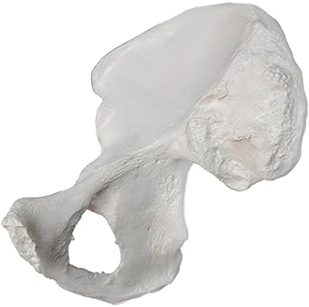 Amazon Com Hip Bone Model Right Anatomically Accurate
 Hip Bones Anatomy Os Coxae Pelvic Girdle Ilium Ischium
 Coxal Bone Landmarks Images Ankle Anatomy Anatomy Bones
 Hip Bone Coxal Bone Pelvic Bone
![]() The Pelvis Human Anatomy And Physiology Lab Bsb 141
The Pelvis Human Anatomy And Physiology Lab Bsb 141
 Anatomy Gross Anatomy Physiology Cells Cytology Cell
 Coxal Bones Quiz By Lolwut77
 Amazon Com Hip Bone Model Left Anatomically Accurate
Hip Bone Coxal Bone Pelvic Bone
 Index 5
The Hip Bone Ilium Ischium Pubis Teachmeanatomy
 The Pelvic Girdle Human Anatomy And Physiology Lab Bsb 141
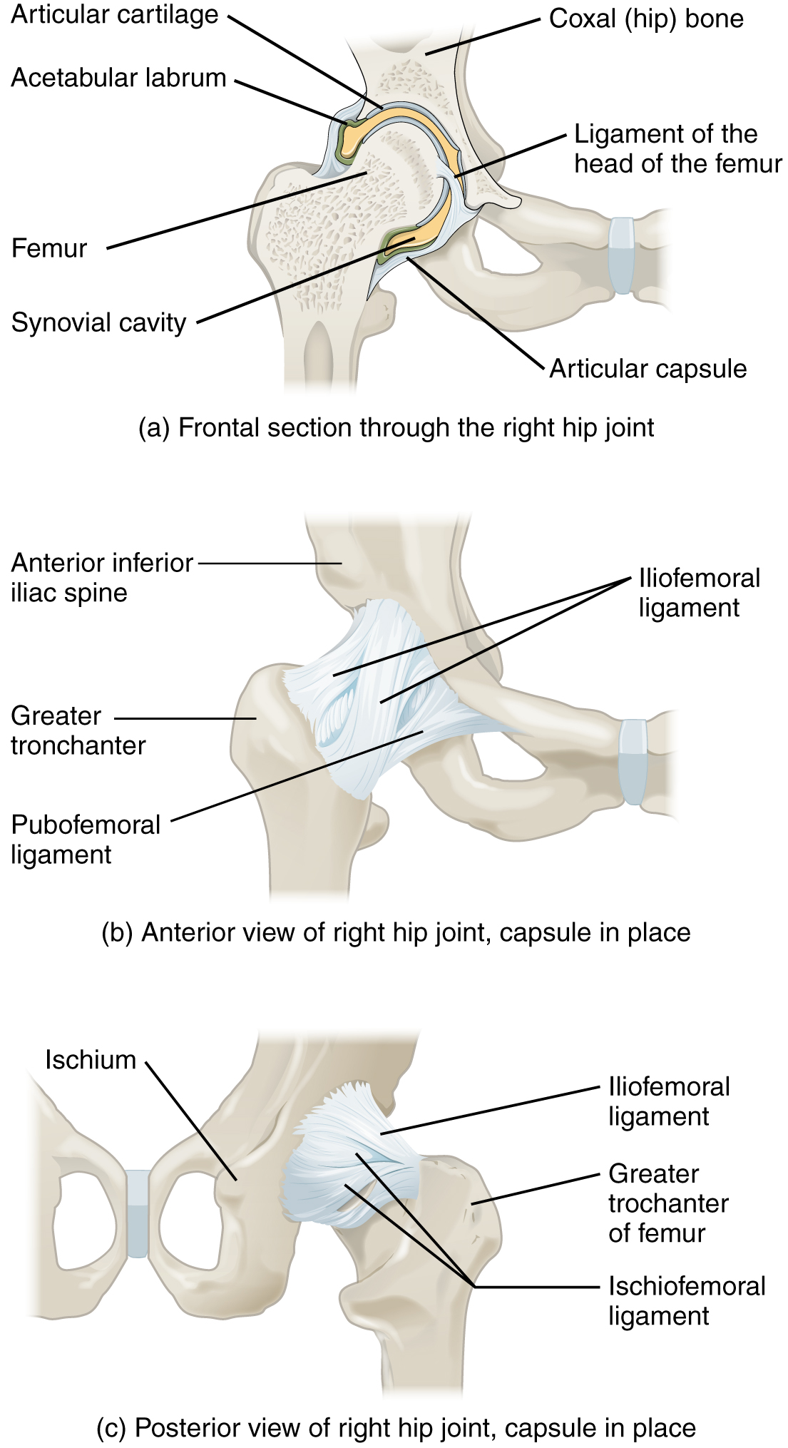 Joints Ligaments And Connective Tissues Advanced Anatomy
 Pelvic Bone Liac Crest Sacroiliac Joint Lliac Fossa Lium
 Medial View Of Coxal Bone Purposegames
Coxal Bone Definition Blog
 Hip Coxal Bone Lateral View Diagram Quizlet
 Hip Bone Pelvic Bone Os Coxa Innominate Bone Or Coxal
 Pelvic Girdle
 Os Coxae Diagram Human Reading Industrial Wiring Diagrams
 Ischium At Odessa College Studyblue
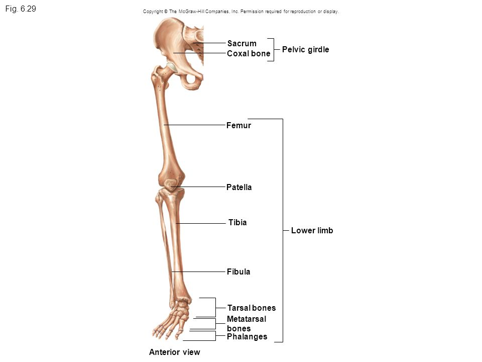 Fig Sacrum Pelvic Girdle Coxal Bone Femur Patella Tibia
 Hip Bone Wikipedia
 Coxal Bone
How To Treat Coxal Bone Pain Quora
 Hip And Femur Skeletal System Portfolio
 Coxal Bone
 Pelvic Girdle Coxal Bones Lower Limb
 Coxal Bone Lateral View Diagram Quizlet
 Pelvic Girdle Coxal Bones Lower Limb
 Coxal Pelvic Bone Posterior View With Labels Appendic
 Hip Bone Wikipedia
 Coxal Bones Flashcards Quizlet
 Hip Bone Anatomy Introduction


Belum ada Komentar untuk "Coxal Bone Anatomy"
Posting Komentar