X Ray Foot Anatomy
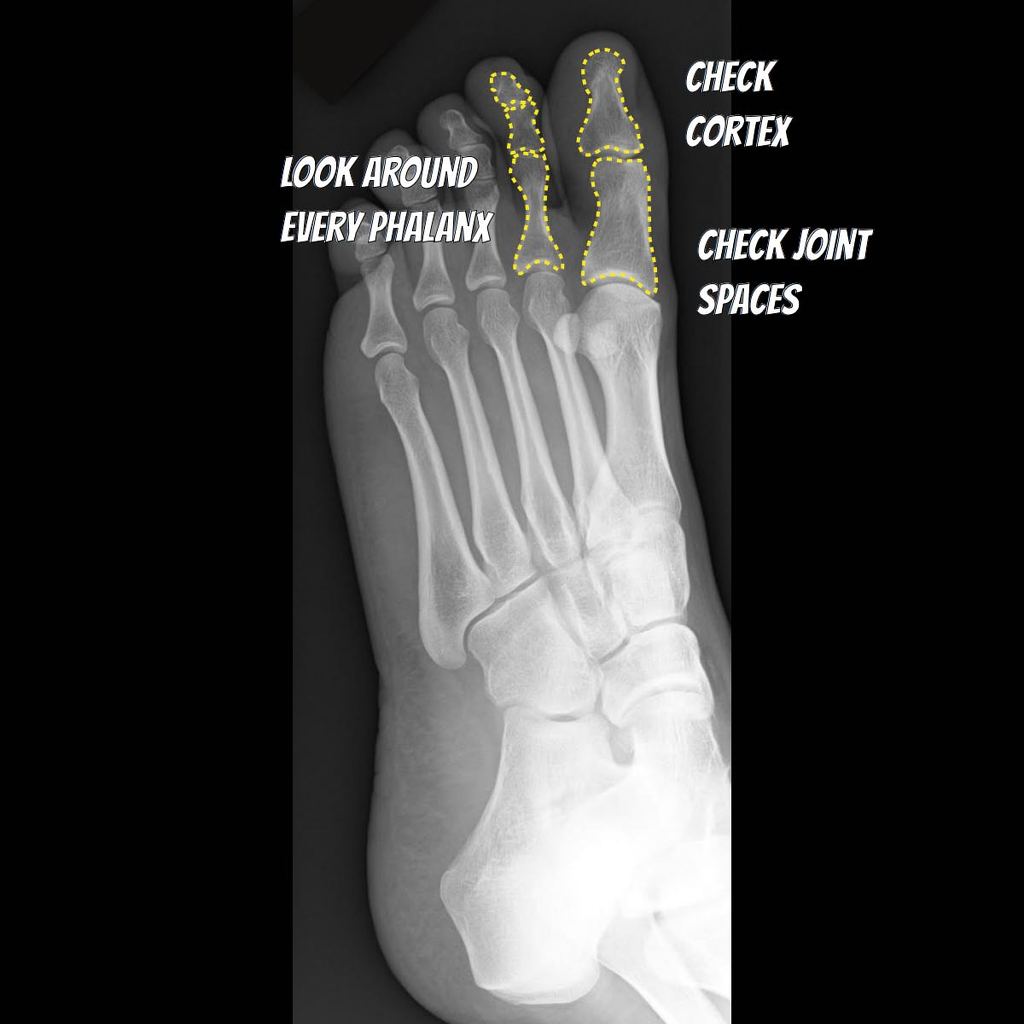
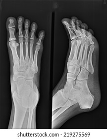
The foot series is comprised of a dorsoplantar dp medial oblique and a lateral projection. When checking any post traumatic foot x ray it is crucial to assess alignment of the bones at the joints.
 Radiology Quiz 29044 Radiopaedia Orgviewing Playlist
For more anatomy content please follow us and visit our website.

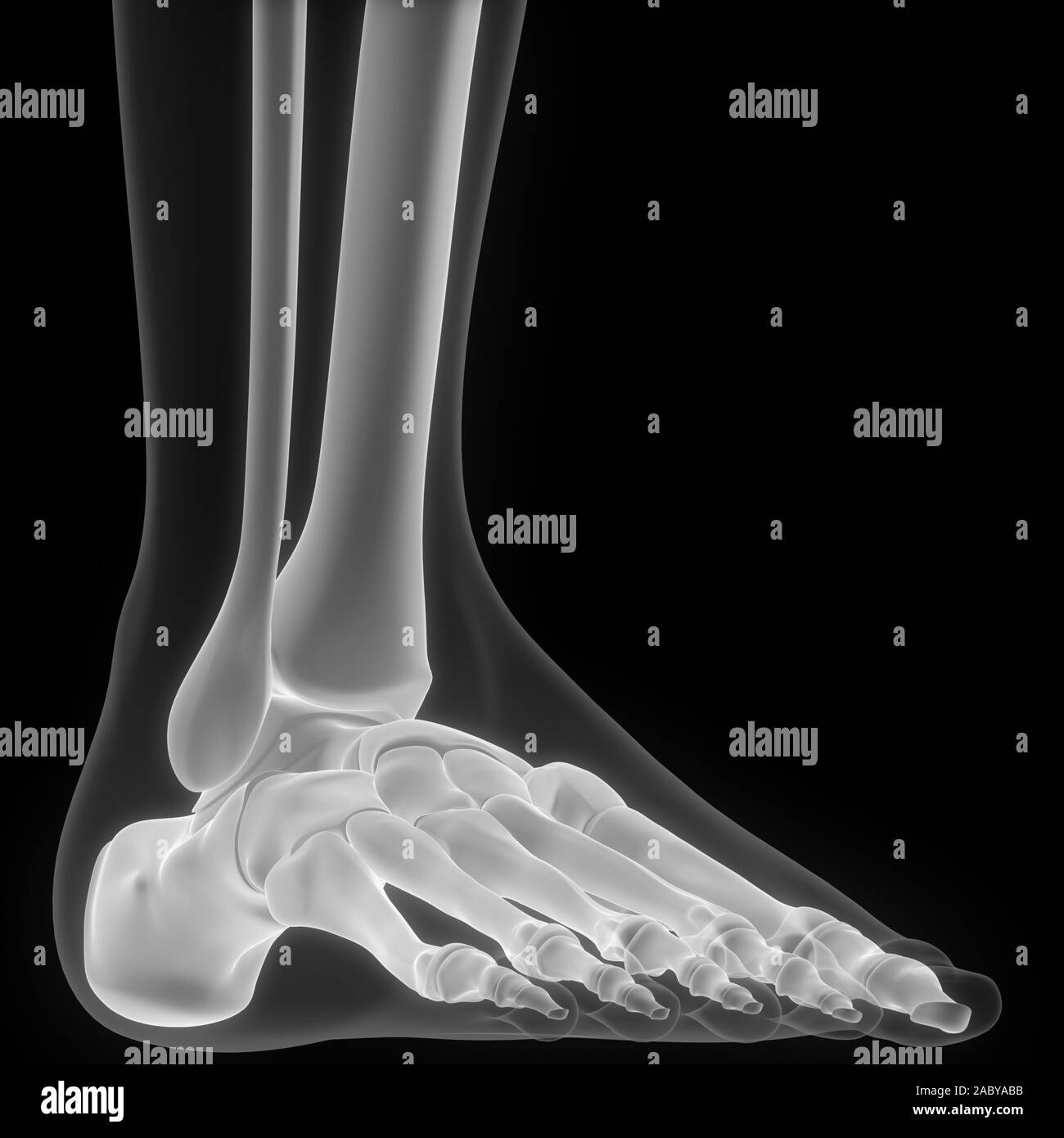
X ray foot anatomy. Approach to foot series. We hope this picture foot ankle x ray lateral view can help you study and research. Remember to check the whole film though.
We think this is the most useful anatomy picture that you need. Normal radiographic anatomy of the foot. This test will be able to show any crack or break in the bones in the ankle 4 9.
This webpage presents the anatomical structures found on foot radiograph. We are pleased to provide you with the picture named foot x ray anatomy. The series is often utilized in emergency departments after trauma or sports related injuries 24.
It is performed to look for evidence of injury or pathology affecting the foot often after trauma. A foot x ray also known as foot series or foot radiograph is a set of two x rays of the foot. There are more than a hundred muscles tendons and ligaments.
It is performed to look for evidence of injury or pathology affecting the foot often after trauma. The human foot has 26 bones and 33 joints. Normal foot and ankle x ray anatomy.
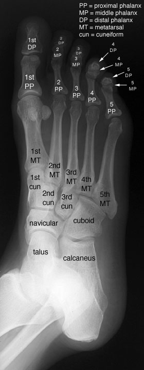
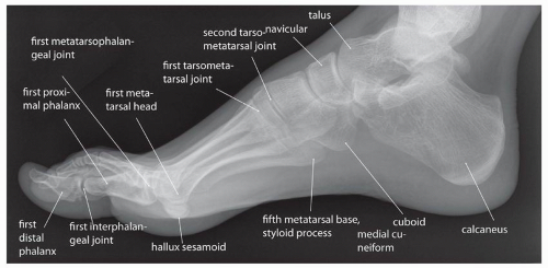
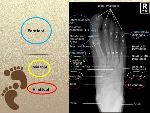
Normal foot and ankle x rays. Foot x ray anatomy in this image you will find distal phalanges interphalangeal joint proximal phalanges metatarso phalangeal joints sesamoid bones metatarsals intermediate phalanges in it. Normal radiographic anatomy of the foot.
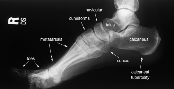
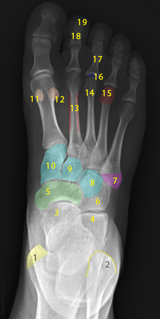
1 calcaneus 2 cuboid. Foot radiographs are commonly performed in emergency departments usually after sport related trauma and often with a clinical request that states lateral border pain. Often a foot x ray is also requested for the investigation of osteomyelitis arthritides or a bone lesion.
1 fibula 2 cuboid 3 5th metatarsal bone 4 tibia 5 talus 6 navicular 7 cuneiform 8 1st metatarsal bone 9 proximal phalanx 10 distal phalanx. The foot is a very stable composition of bones supported by strong ligaments. This is a very complex structure due to the need to support your entire body weight.
Normal radiographic anatomy of the foot. The x ray is the most commonly requested radiographic examination because of its availability. Detailed anatomy description of foot.
Loss of joint alignment can represent severe injury even in the absence of a fracture.
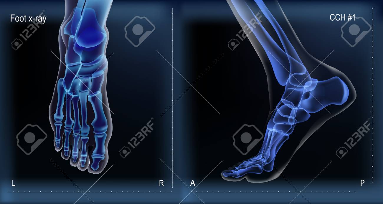 Dark Navy Blue Vector Realistic Medial And Top X Ray Of Skeleton
 X Ray Of Human Leg Bone Posterior View Red Highlights In
 Foot Ankle X Ray Lateral View
Normal Radiographic Anatomy Of The Ankle Radiology Case
 Anatomy Of The Bones Of The Foot The Bmj
 Human Skeleton System Bone Joints Anatomy X Ray 3d Rendering
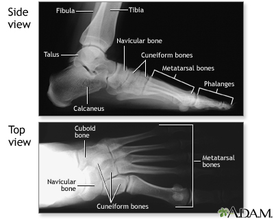 Normal Foot X Ray Medlineplus Medical Encyclopedia Image
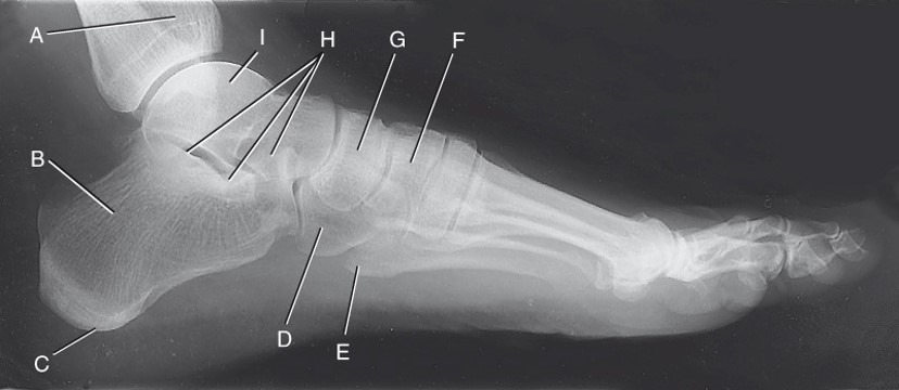 Diagram Foot Anatomy X Ray Lateral View Diagram Quizlet
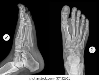
 Oblique And Anterior Posterior View X Rays Of A Normal Foot
 Flat Feet Wikipedia
 Pin On Bones
 Anatomy Of Foot Stock Photos Images Photography


 Foot Anatomy Canvas Prints Fine Art America
 Game Statistics 3 View Foot X Ray Anatomy Purposegames
 X Ray Foot Findings Fracture Stock Image Image Of Care
 X Ray Of Foot Ankle Wall Mural Anatomy Wallpaper Murals
 Free Art Print Of X Ray Foot Illustration
 Diagnostic Imaging Techniques Of The Foot And Ankle
 Normal Radiographic Anatomy Of The Foot Radiology Case
 Foot Annotated X Ray Radiology Case Radiopaedia Org
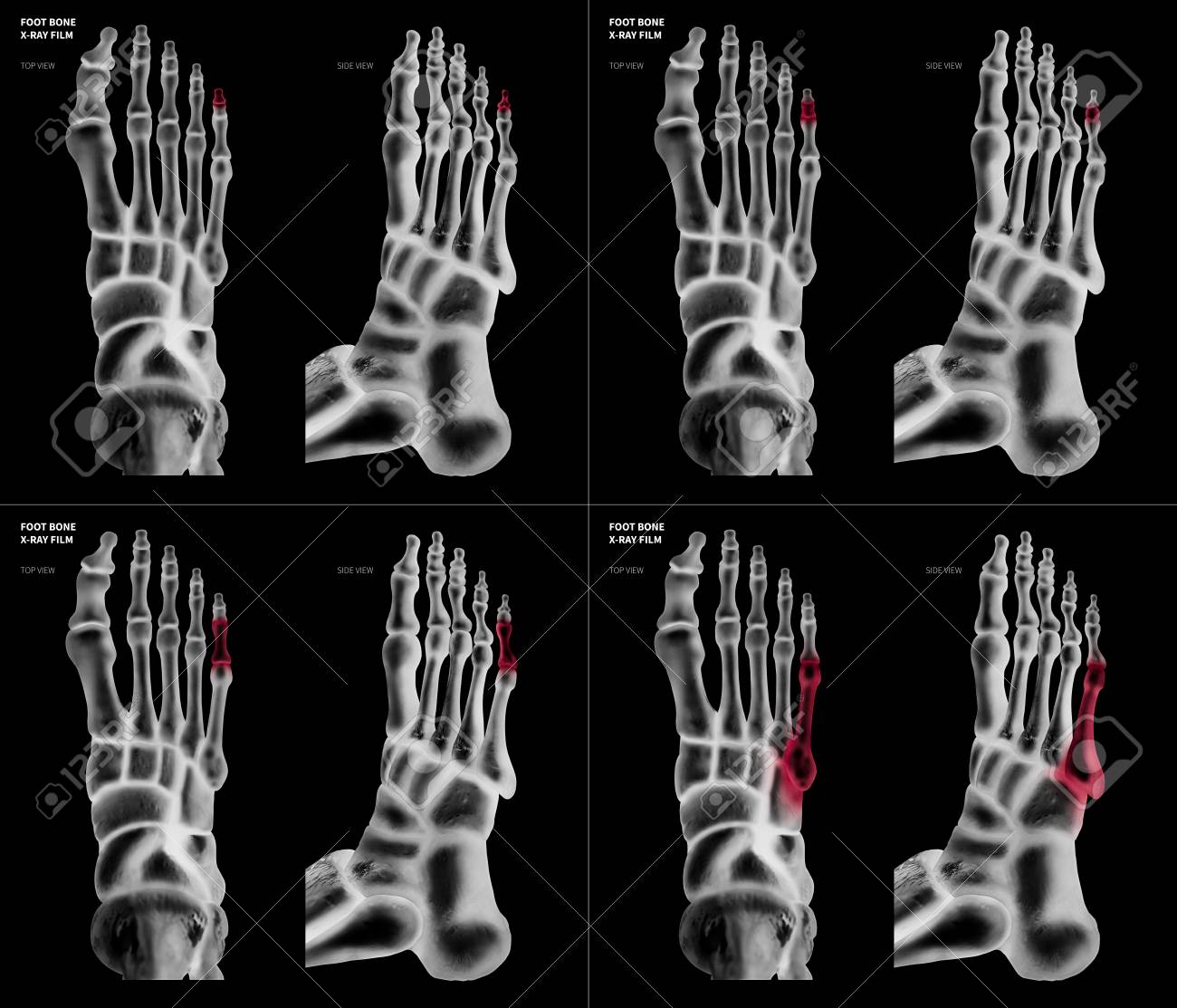 X Ray Film Collection Of Little Toe Foot Bone With Red Highlights
 Royalty Free Foot Xray Stock Images Photos Vectors
 Infographic Diagram Of Human Foot Bone Anatomy System
 X Ray Of Foot Stock Photo Image Of Patient Life Anatomy
 Foot Radiological Anatomy Shorouk Zaki
 X Ray Foot Ap Obl Anatomy And Physiology Part 19
 Science Source Foot Anatomy X Ray
Belum ada Komentar untuk "X Ray Foot Anatomy"
Posting Komentar