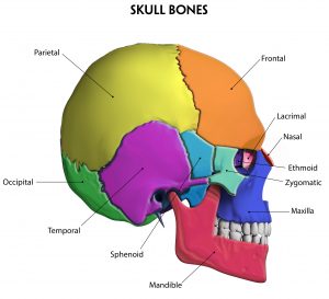
Eye Bone Anatomy
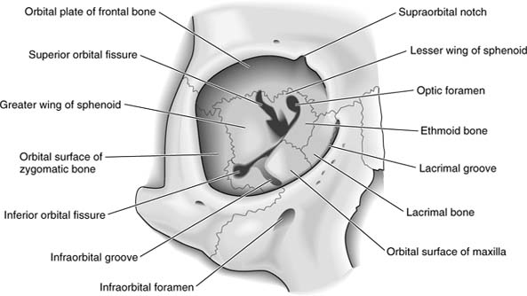
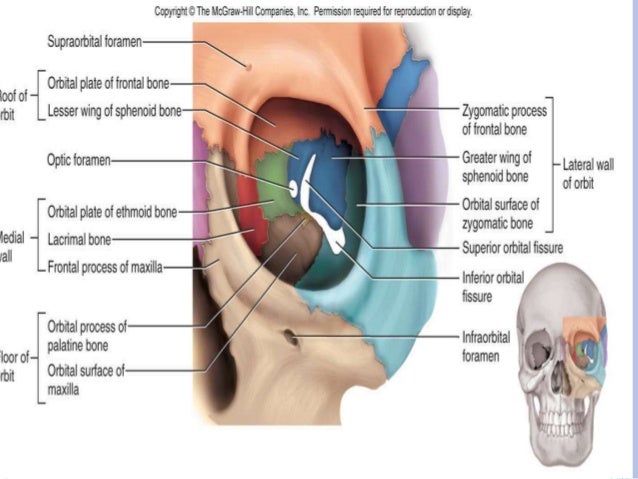
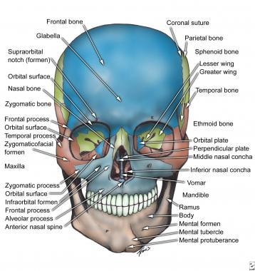
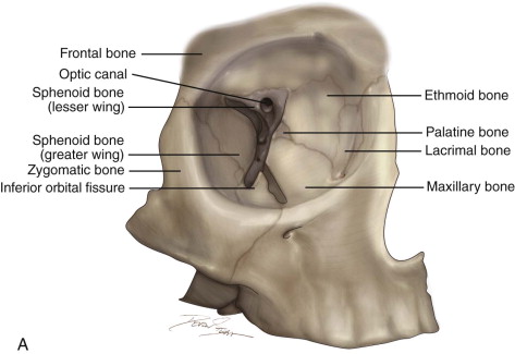
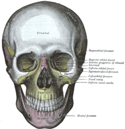
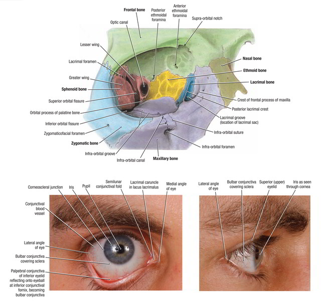
In the adult human the volume of the orbit is 30 millilitres 106 imp fl oz. Lateral wall formed by the zygomatic bone and greater wing of the sphenoid.
 Maxilla Vertebrate Anatomy Britannica
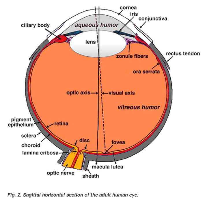
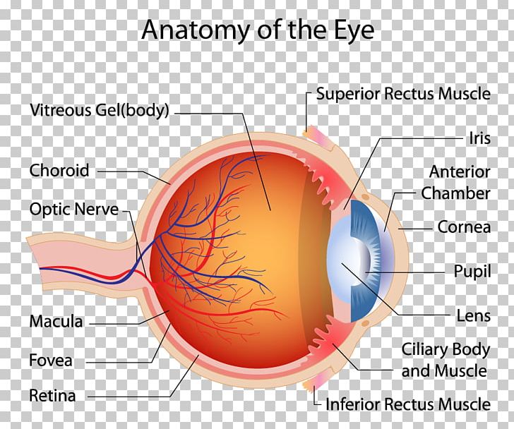
Just behind the iris and pupil lies the lens which helps focus light on the back of your eye.

Eye bone anatomy. It is also known as the orbital rim. Science quiz human eye anatomy random science or biology quiz can you locate the parts of the human eye. Anatomy and physiology of the eye conjunctiva.
Dry eye retinal detachment macular degeneration diabetic retinopathy eye trauma etc. Bones of the skeleton. For ophthalmologists optometrists medical dental and optometry students eye anatomy forms the basis for eye pathology in diseases.
101 us fl oz. The ethmoid bone separates the orbit from the ethmoid sinus. Light projects through your pupil and.
The cornea is the transparent clear layer at the front and. If you have found anatomyzone useful and you would like to support our aim of providing the best free online anatomy resource please consider using the form below to make a contribution towards our development. Things our bodies do.
Orbit bones eye. The conjunctiva is a thin transparent layer of tissues covering the front of the eye. The human eye is an organ which reacts to light and pressure.
As a sense organ the eye allows vision. A closer look at the parts of the eye by liz segre when surveyed about the five senses sight hearing taste smell and touch people consistently report that their eyesight is the mode of perception they value and fear losing most. Here is a fun way mnemonic for learning the bones in the eye socket of the human skull.
Hide this ad show comments. Use this anatomy quiz to learn to locate its components. Most of the eye is filled with a clear gel called the vitreous.
Base opens out into the face and is bounded by the eyelids. Medial wall formed by the ethmoid maxilla lacrimal and sphenoid bones. Orbit anatomy in anatomy the orbit is the cavity or socket of the skull in which the eye and its appendages are situated.
Anatomy of the human eye anatomy of the eye includes lacrimal gland cornea conjunctiva uvea iris choroid ciliary body lens blood supply retina vitreous optic nerve. Test your knowledge on this science quiz to see how you do and compare your score to others. Orbit can refer to the bony socket or it can also be used to imply the contents.
Apex located at the opening to the optic canal the optic foramen. Can you locate the parts of the human eye. The white part of the eye that one sees when looking at oneself in the mirror is.
 Dark Circles Remedies Treatment Without Surgery In Paris
 Orbital Bone Anatomy Eye Anatomy Facial Anatomy
 Laminated Eye Anatomical Poster Human Eye Anatomy Chart 18 X 27
 Benefits Of Foot Zoning The Cranial Bones
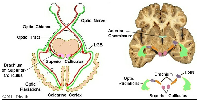 Neuroanatomy Online Lab 7 Visual System Gross Anatomy
 Eye Anatomy And Vision Course Hero
 Instant Anatomy Head And Neck Areas Organs Eye Orbit
 Orbit In Cross Section Anatomy The Eyes Have It
 Osteopathy St Olav Eye Clinic
 Gross Anatomy Of The Eye By Helga Kolb Webvision
 Orbital Bones Ophthalmology Review
 Human Eye Ball Anatomy Physiology Diagram
 Human Eye Macula Of Retina Muscle Anatomy Png Clipart
 Anatomy Of The Orbit
 Jaw Bones Diagram Front Reading Industrial Wiring Diagrams
 Orbital Bones Ophthalmology Review
 The Orbit And Eye Pocket Dentistry
 Retina Macula Lutea Choroid Anatomy Eyeball Anatomy Eye
 Infraorbital Foramen Wikipedia
 Easy Notes On Eyelids Learn In Just 4 Minutes Earth S Lab
 The Frontal Bone John Hawks Weblog
 Skull Bones Of The Orbit Human Anatomy Kenhub
 Ocular Anatomy Columbia Asia Hospital Malaysia
 Eye Orbit Bones Above 3d Human Anatomy Organs


Belum ada Komentar untuk "Eye Bone Anatomy"
Posting Komentar