Ultrasound Anatomy
Description examining your growing babybabies. In special cases a detailed examination of the fetal heart and.
 Essential Ultrasound Anatomy
Heart rate rhythm 4 chamber views.
Ultrasound anatomy. Liver ultrasound showing education liver segments normal liver anatomy portal vein hepatic veins the biliary tree and ultrasound scanning protocol worksheets. Most anatomy scans are performed in the second trimester of pregnancy typically at 20 weeks but they can be done anytime between 18 weeks and 22 weeks. Otero and diego a.
Manual of small animal regional anesthesia. The purpose of the 20 to 22 week ultrasound is to look at all of the fetuss anatomy and to determine if all looks normal. There are two main types of fetal ultrasound exams.
A transabdominal fetal ultrasound is done by moving a transducer. The second trimester extends from 13 weeks and 0 days to 27 weeks and 6 days of gestation although the majority of these studies are performed between 18 and 23 weeks. Those who want to can find out the sex of the baby if desired.
Things that cant be seen in earlier scans such as spinal cord. Ultrasound evaluation of normal fetal anatomy. During the scan we will examine each part of the fetal body in detail.
Instrumentation has improved steadily yielding both improved and more consistent image quality. Skull shape integrity bpd and hc measurements. Brain ventricles choroid plexus mid brain posterior fossa cerebellum cisterna magna.
With this type of fetal ultrasound a wandlike device called. The anatomy scan is a level 2 ultrasound which is typically performed on pregnant women between 18 and 22 weeks. The gender of your babybabies can usually be determined at this ultrasound.
Neck nuchal fold thickness. Our understanding of normal fetal anatomy as seen on sonograms continues to be an area of considerable growth. The following fetal parts are checked during the anatomy ultrasound.
Illustrated anatomy for nerve stimulation and ultrasound guided nerve blocks by pablo e. If you have a condition that needs to be monitored such as carrying multiples you may have more than one detailed ultrasound. Portela jul 1 2019 hardcover.
When a level 2 ultrasound is done. The second trimester scan is a routine ultrasound examination in many countries that is primarily used to assess fetal anatomy and detect the presence of any fetal anomalies.
 Abdominal Ultrasound Registry Review
 Ten Questions Every Pregnant Woman Should Ask
 Ultrasound Of The Pancreas What Normal Looks Like
 Point Of Care Ultrasound Complete Anatomy
 Atlas Of Fetal Sectional Anatomy With Ultrasound And Magnetic Resonance Imaging
 Ultrasound Anatomy Of Sciatic Nerve Sciatic N Block
 Presentation1 Abdominal Ultrasound Anatomy
 Ultrasound Anatomy Ultrasound Anatomy Derick Mussen
 Msk Ultrasound On Twitter Confused About The Anatomy Of
 Anatomy Ultrasound December 2018 Babies Forums What To
 Liver Anatomy And Segments By Ultrasound In Arabic
 Startradiology
 Neonatal Cranial Ultrasound Standard Views
 Anomaly Scan Wikipedia
 Shelby S 24 Week Ultrasound Anatomy Scan Mommymandy L
 Normal Anatomy Of Gall Bladder Ultrasound Sonography
 Shoulder Ultrasound Anatomy Qir Basic Msus Anatom Yof Th
 Startradiology
 Ultrasound Guided Rectus Sheath Block Hadzic S Peripheral
 20 Week Anatomy Ultrasound Youtube
 Prenatal Ultrasounds Columbia Shores Obgyn
 Liver Hilum Anatomy Ultrasound
 The 20 Week Ultrasound Anatomy Scan Awesome First Sights
 Emergency Ultrasound
 Liver Ultrasound Ultrasound Sonography Vascular
 Liver Measurement Ultrasound Pancreas And Its Proportions
 Kidney Ultrasound Anatomy And Scanning Methods Abdominal Key
 1 Normal Ultrasound Anatomy Seen Above The Asis Download
 Normal Ultrasound Anatomy Of The Eye In The Correct Plane
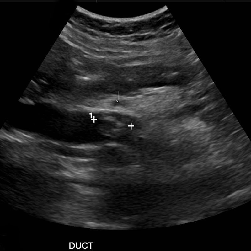
Cbd Ultrasound Anatomy Biliary Ultrasound 2019 10 06


Belum ada Komentar untuk "Ultrasound Anatomy"
Posting Komentar