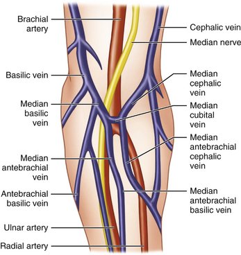
Vein Anatomy Arm
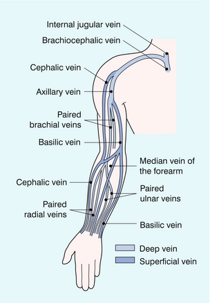
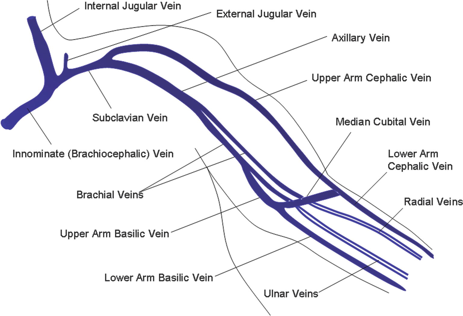
As you reach the proximal arm the axillary vein will divide into the basilic and brachial veins. The veins return oxygen.
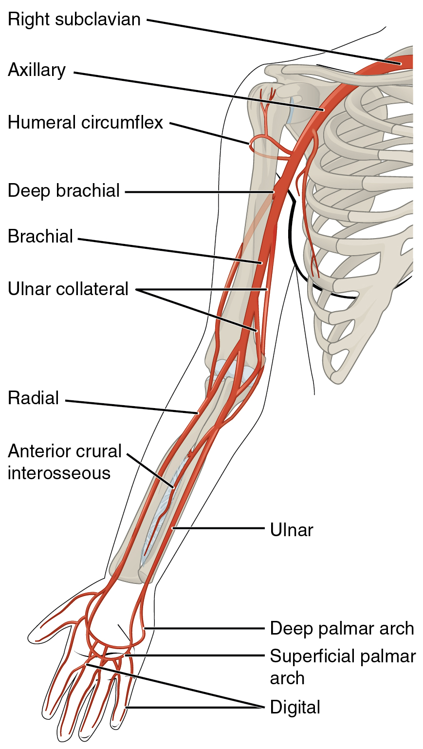 20 5 Circulatory Pathways Anatomy And Physiology
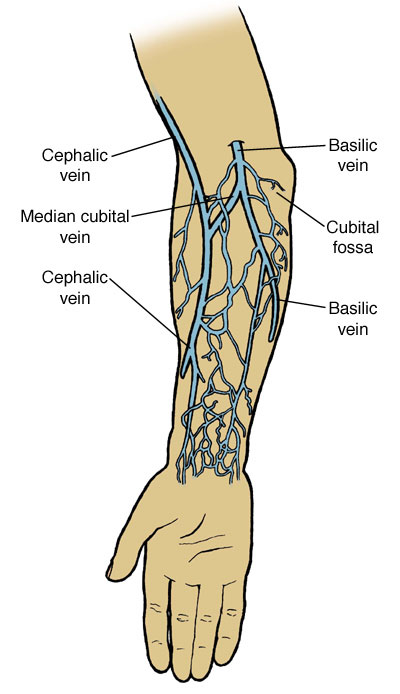
Making its way through the basilic vein is the median basilica vein located in the lower part of the elbow which works as a communicator in the arm.

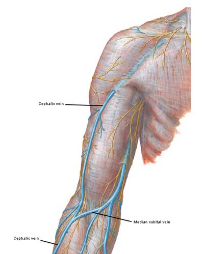
Vein anatomy arm. From the radial side lateral side of the forearm the cephalic vein runs up from the anatomical snuffbox along the preaxial border of the upper limb. The veins of the arm may be divided into two groups. Muscular contractions press on the veins and form a very effective mechanism of venous return.
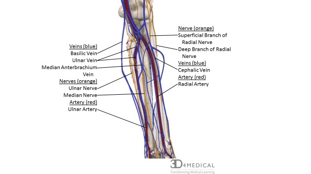
The basilic vein originates from the dorsal venous network of the hand and ascends the medial aspect of the upper limb. Upper arm veins brachial basilic the basilic vein is the larger and is more superficial. Continue from the axillary vein checking in transverse that the basilic and brachial veins of the upper arm are compressible.
Deep veins and superficial veins. The cephalic and basilic veins are major superficial veins of the forearm. This becomes still more effective.
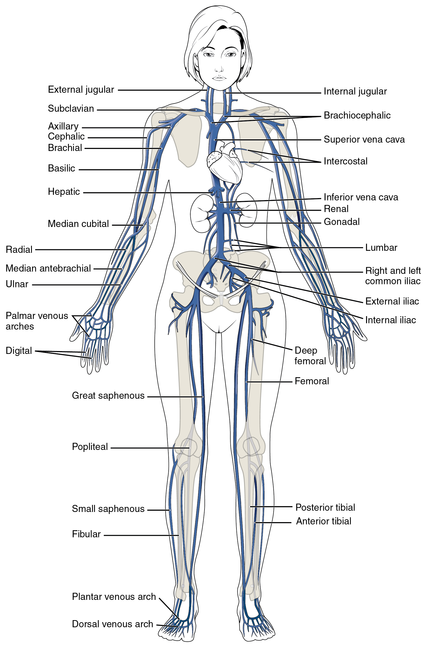
The arteries deliver freshly oxygenated blood to muscles and bone. They are located within the subcutaneous tissue of the upper limb. The major superficial veins of the upper limb are the cephalic and basilic veins.
At the border of the teres major the vein moves deep into the arm. Vein problems are typically the result of a blockage or defect. The vessels of the arms are part of the circulatory system which provides nutrients to the tissues.
Continuing upwards through the forward part of the elbow the cephalic vein makes its way through the valley created by the biceps brachii and the brachioradialis on either side of it. Usually single but may be duplicated. Blockages occur due to blood clots that develop in either superficial veins or deep veins most often in the legs or arms.
The deep veins for the most part accompany arteries within the fascial sleeve whereas the superficial veins lie for most of their course outside the fascial sleeve. Within the tight sleeve of the deep fascia as is seen in the lower limbs. Blood clots develop when blood cells known as platelets or thrombocytes become activated due to a vein injury or disorder.
How Many Veins Are In The Human Body Quora
 Veins Arm Stock Photos Veins Arm Stock Images Alamy
 Frank Netter Page 8 Outlander Anatomy
 Vein Pelvis Abdomen Human Anatomy Arm Transparent
 Nerves Blood Vessels And Lymph Advanced Anatomy 2nd Ed
 Arm Vein Anatomy Diagram Quizlet
 Upper Limb Surface Anatomy Trapezius Muscle Cephalic Vein
![]() Cephalic Vein Anatomy And Clinical Points Kenhub
Cephalic Vein Anatomy And Clinical Points Kenhub
 The Peripheral Veins Clinical Gate

 Superior Vena Cava And Right Arm Vein Anatomy
 Vein Wikipedia
 Median Cubital Antebrachial Veins Locations Functions
 Vein Arm Anatomy Stock Illustrations 226 Vein Arm Anatomy
 Upper Limb Anatomy
 Chapter 33 Venous And Intraosseous Access In Adults

![]() Cephalic Vein Anatomy And Clinical Points Kenhub
Cephalic Vein Anatomy And Clinical Points Kenhub
 Cephalic Vein Wikipedia
 Venous Drainage Of The Lower Limb Teachmeanatomy
 Thumb Elbow Arm Anatomy Vein Png 600x600px Watercolor
 Basilic Vein An Overview Sciencedirect Topics
 Vein Pelvis Forearm Anatomy Arm
 Anatomy Of Gsv And Ssv With Common Variants Of Ssv Gsv
 Anatomy Atlases Anatomy Of First Aid A Case Study Approach
 Upper Extremity Venous Thrombosis Thoracic Key
 Clinical Anatomy Of The Cephalic Vein For Safe Performance
![]() Veins Of The Upper Limb Anatomy Kenhub
Veins Of The Upper Limb Anatomy Kenhub
 Venipuncture Part 1 Anatomy Of The Arm And Vein Location
 20 5 Circulatory Pathways Anatomy And Physiology
 23 Anatomy For Venipuncture Pocket Dentistry

Belum ada Komentar untuk "Vein Anatomy Arm"
Posting Komentar