Fetal Heart Anatomy
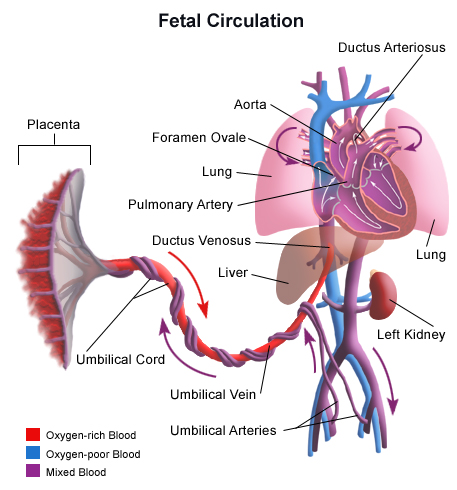
Normal 3 vessel view in a 28 week fetus. Fetal circulation after birth the circulation is divided into two separate sides that are not connected.
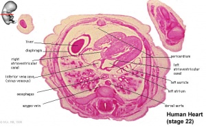 Cardiovascular System Heart Development Embryology
Contents of this page.

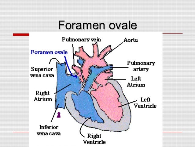
Fetal heart anatomy. Normal sonographic anatomy of the fetal heart. The left side of the heart consists of the pulmonary veins left atrium left ventricle and aorta. The majority of the babies with chd are born to parents with no identifiable risk factors.
Ultrasound image of the right ventricular outflow tract rvot ultrasound images of the lvot left ventricular outflow tract some more images of lvot and rvot. The fetus is supine the left atrium is nearest to the spine and the apex of the heart points to the left. Fetal cardiac examination is performed either as a part of screening fetal sonography for a low risk population or as a complete diagnostic test for groups at high risk for chd table 14 1.
Confirm that the heart is on the fetal left. Start a free trial of quizlet plus by thanksgiving lock in 50 off all year try it free ends in 02d 05h 28m 23s. The right side of the heart consists of the superior and inferior vena cava the right atrium the right ventricle.
Choose from 500 different sets of fetal heart anatomy flashcards on quizlet. Fetal heart ultrasouns of four chamber view. The fetus is in supine position and the border of the right ventricle is under the sternum.
The left ventricle occupies most of the left cardiac border. In terms of its size the normal heart occupies about 13 of the fetal thorax and its transverse diameter of the heart must not exceed half of the transverse diameter of the chest figure 1. Normal fetal cardiac anatomy so as to assess and diagnose the many and varied congenital cardiac malformations it has long been recognized that the normal heart can best be assessed in terms of three so called segments.
The heart should be angled 45degrees to the left and occupy approximately 13 of the chest. Mere presence of the heart in the location as described above and the finding of normal arrangement of the remaining organs however does not guarantee cardiac normality.
 Fetal Heart Rate Tracing Physiology Fetal Heart Rate
 Advanced Evaluation Of The Fetal Heart
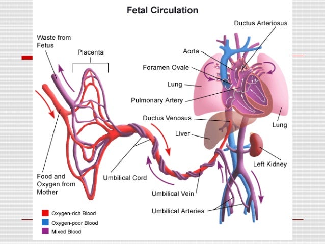 Fetal Heart Circulation
 Key Screening Views Of The Fetal Heart Part 3 4 Chamber View
 Development Of Blood Vessels And Fetal Circulation Anatomy

 Fetus Circulation An Overview Sciencedirect Topics


 Fetal Circulation Heart Circulation Medical Anatomy
 Blood Circulation In The Fetus And Newborn Children S
 6 Fetal Heart Anatomy And Electrical Activation Sequence
Heart Anatomy Chambers Valves And Vessels Anatomy
 Anatomy Of The Normal Fetal Heart The Basis For

 Hypoplastic Left Heart Syndrome Symptoms And Causes Mayo
 Fetal Circulation And Perinatal Programming Esc Cardiomed
 6 Fetal Heart Anatomy And Electrical Activation Sequence
 Atrium Heart Wikipedia
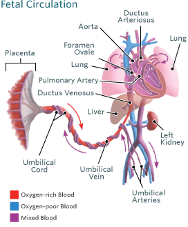
 Fetal Heart Circulation
 Pacific Medical Training Acls Bls Pals Certification
 Congenital Heart Defect Wikipedia
![]() Ductus Arteriosus Anatomy And Explanation Kenhub
Ductus Arteriosus Anatomy And Explanation Kenhub
 Pda Pfo And Fetal Circulation
Belum ada Komentar untuk "Fetal Heart Anatomy"
Posting Komentar