Anatomy Of Bone
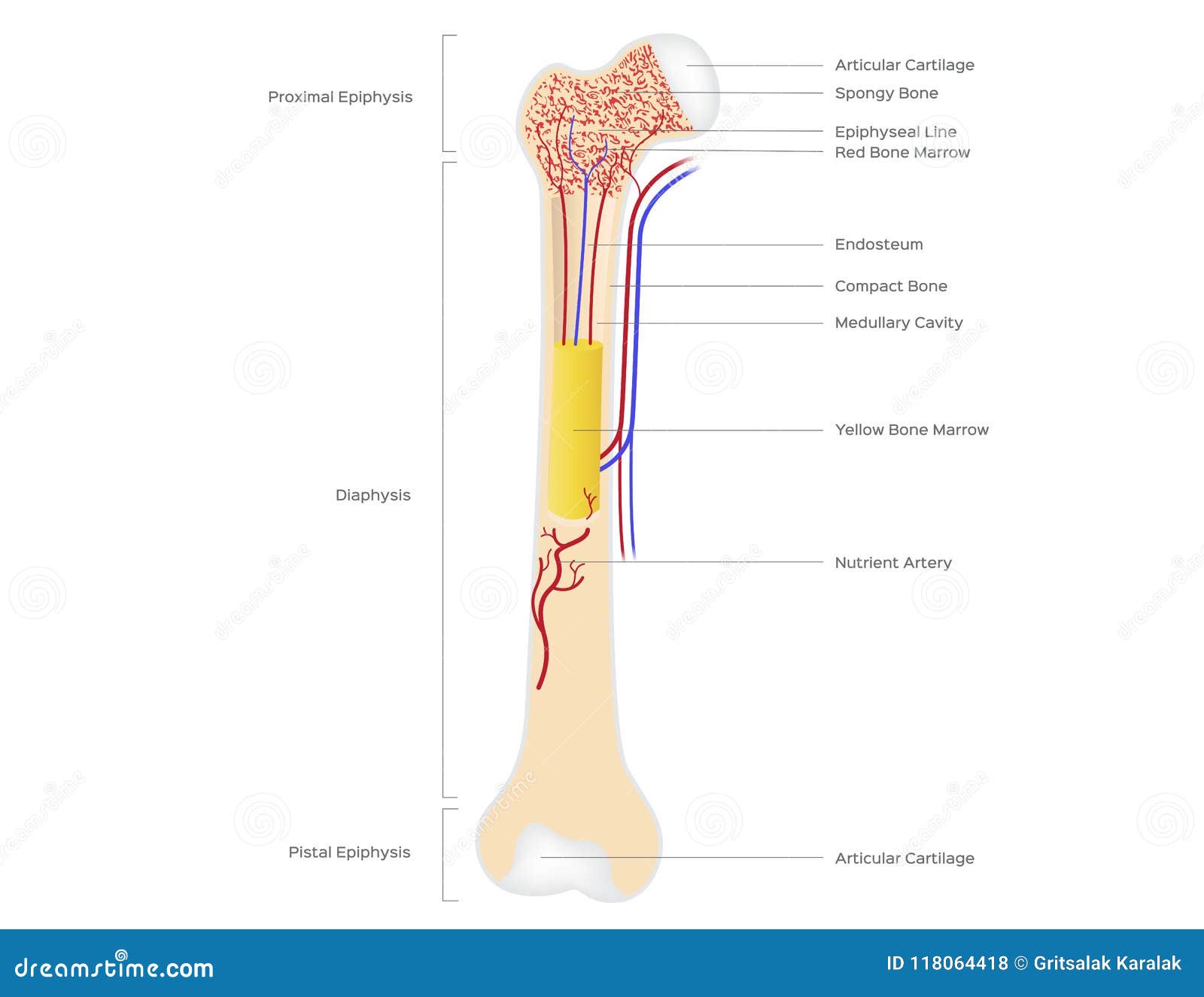
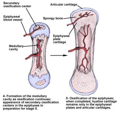
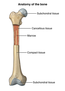
A long bone has two parts. The structure of a long bone allows for the best visualization of all of the parts of a bone figure 1.
 Anatomy Of The Bones In Your Body Skeleton Bones Human
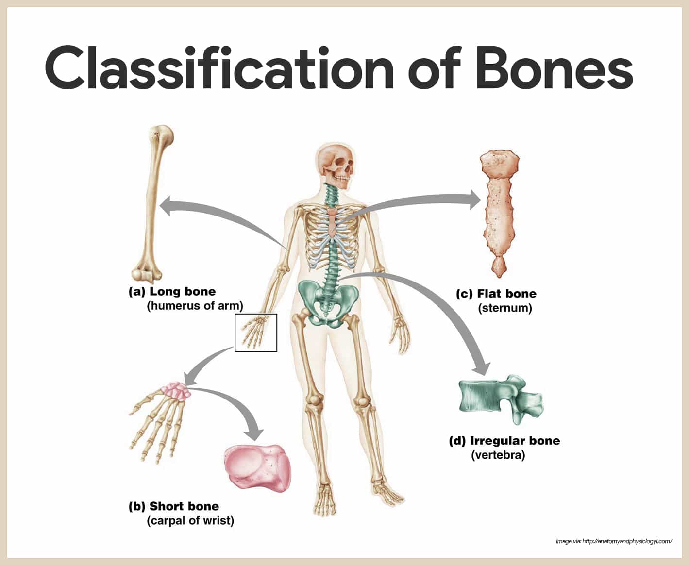
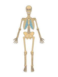
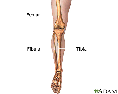
Long bones include the clavicles humeri radii ulnae metacarpals femurs tibiae fibulae metatarsals and phalanges.
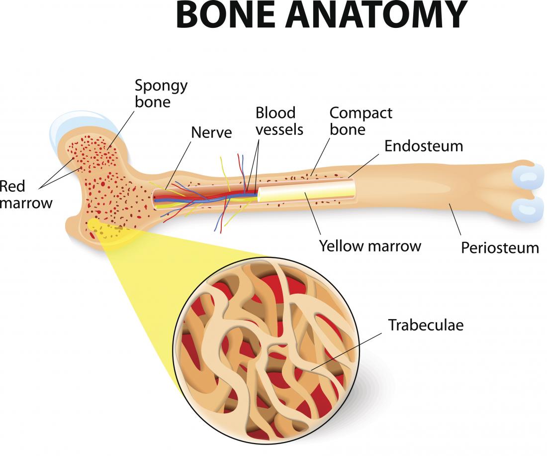
Anatomy of bone. The inside of your bones are filled with a soft tissue called marrow. He talks about the anatomy of the skeletal system including the flat short and irregular bones and their individual arrangements of compact and spongy bone. Gross anatomy of bone.
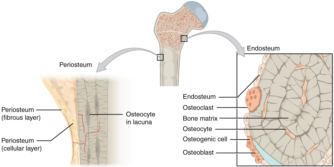
Tendons and ligaments also attach to bones at the periosteum. Gross anatomy of bone. The diaphysis and the epiphysis.
There are two types of bone marrow. Flat bones are thin and generally curved with two parallel layers of compact bones sandwiching. Anatomy of the typical long bone as well as microscopic anatomy of bone tissue.
Overall the bones of the body are an organ made up of bone tissue bone marrow blood vessels epithelium and nerves. Short bones are roughly cube shaped and have only a thin layer of compact bone surrounding. Platelets are small pieces of cells that help you stop bleeding when you get a cut.
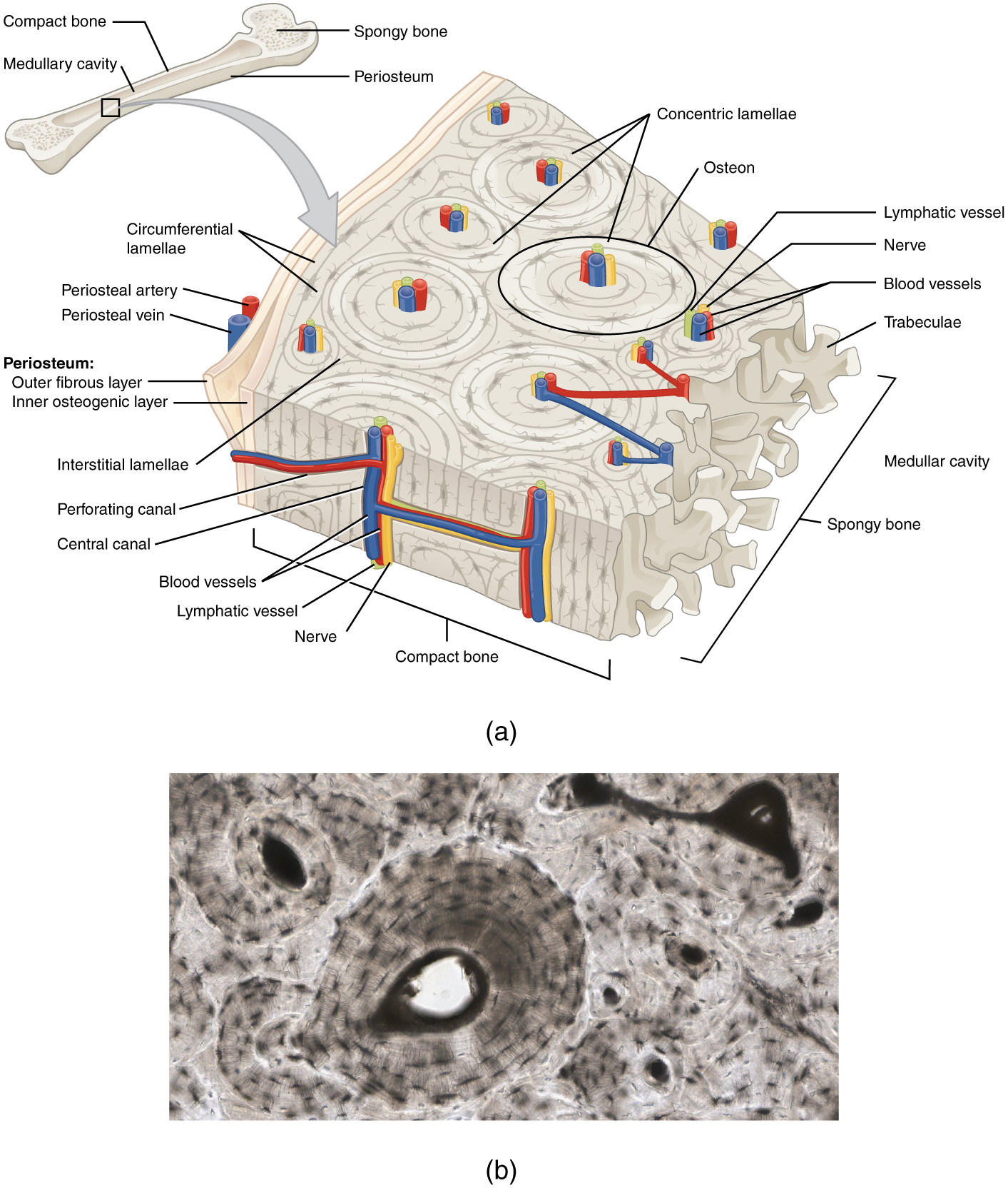
The periosteum contains blood vessels nerves and lymphatic vessels that nourish compact bone. The two principal components of this material collagen and calcium phosphate distinguish bone from such other hard tissues as chitin enamel and shell. Bone is made of bone tissue a type of dense connective tissue.
Bone osseous tissue is the structural and supportive connective tissue of the body that forms the rigid part of the bones that make up the skeleton. Red bone marrow is where red blood cells are formed. The four general categories of bones are long bones short bones flat bones and irregular bones.
All bones contain bone marrow which is either red or yellow. Red bone marrow is where all new red blood cells white blood cells and platelets are made. Types long bones are characterized by a shaft the diaphysis that is much longer than its width.
The diaphysis is the tubular shaft that runs between the proximal and distal ends of the bone. Anatomy bone rigid body tissue consisting of cells embedded in an abundant hard intercellular material. The outer surface of the bone is covered with a fibrous membrane called the periosteum peri around or surrounding.
Yellow bone marrow is made up of fat or adipose cells.
 The Upper Limbs Human Anatomy And Physiology Lab Bsb 141
 Diagram Of Human Bone Anatomy Useful For Education In Schools
 Femur Bone Anatomy Landmarks And Muscle Attachments
 Skeletal System Anatomy And Physiology Nurseslabs
 6 3 Bone Structure Anatomy And Physiology
 6 3 Bone Structure Anatomy And Physiology
 Bone Structure Anatomy For Science And Education
 The Human Skeleton Laminated Anatomy Chart Skeleton
 Osteology Bone Anatomy Overview Gross Anatomy Overview
 Human Bone Anatomy And Physiology Osteology
 Skeletal System Anatomy And Physiology Nurseslabs
 Anatomy Specific Bones Of The Feet
 Introduction To Bone Boundless Anatomy And Physiology
 Bones Types Structure And Function
 Skeletal System Labeled Diagrams Of The Human Skeleton
 Skeletal System Anatomy And Physiology
 Skeletal System Anatomy Function
 Leg Skeletal Anatomy Medlineplus Medical Encyclopedia Image

 Bones Of The Pelvis Hip Bones Anatomy Tutorial
 Skeletal System Anatomical Chart Laminated Human Skeleton Poster
Anatomy 101 Finger Bones The Handcare Blog
 Bone Structure And Function Human Anatomy
 Axis Scientific Human Skeleton Model Anatomy Bundle 5 6 Life Size Skeletal System 206 Bones Interactive Medical Replica 3 Year Warranty Study
Microscopic Anatomy Of Bone Course Hero


Belum ada Komentar untuk "Anatomy Of Bone"
Posting Komentar