X Ray Hand Anatomy
Hand radiograph an approach hand series. Give it a try.
 Skeletal Anatomy 4 And An X Ray Image Of A Hand 5
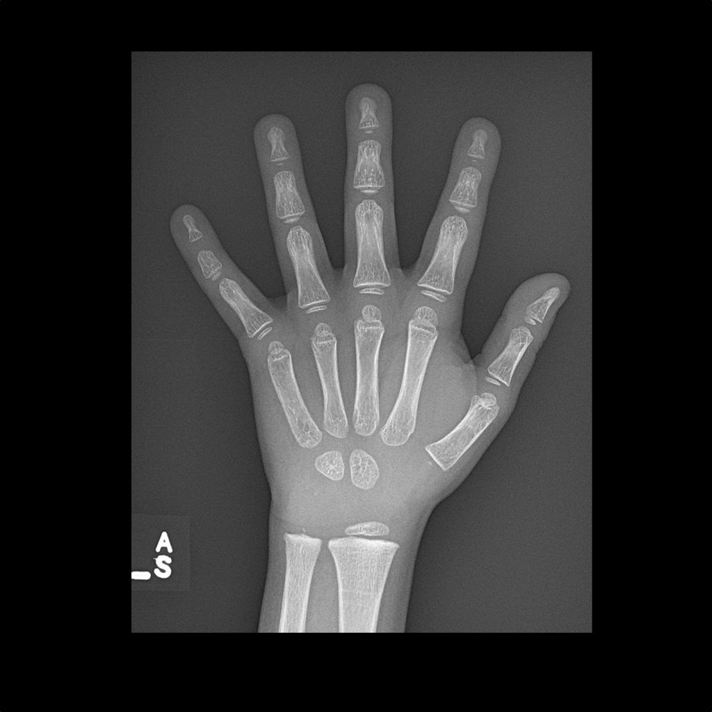
Characteristics of a normal handfinger x ray.

X ray hand anatomy. 3 article feature images from this case. Anatomy of the hand and wrist. X ray games anatomy upper limb pictures quiz.
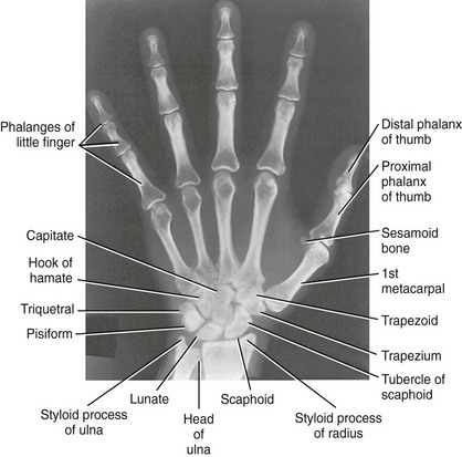
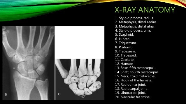
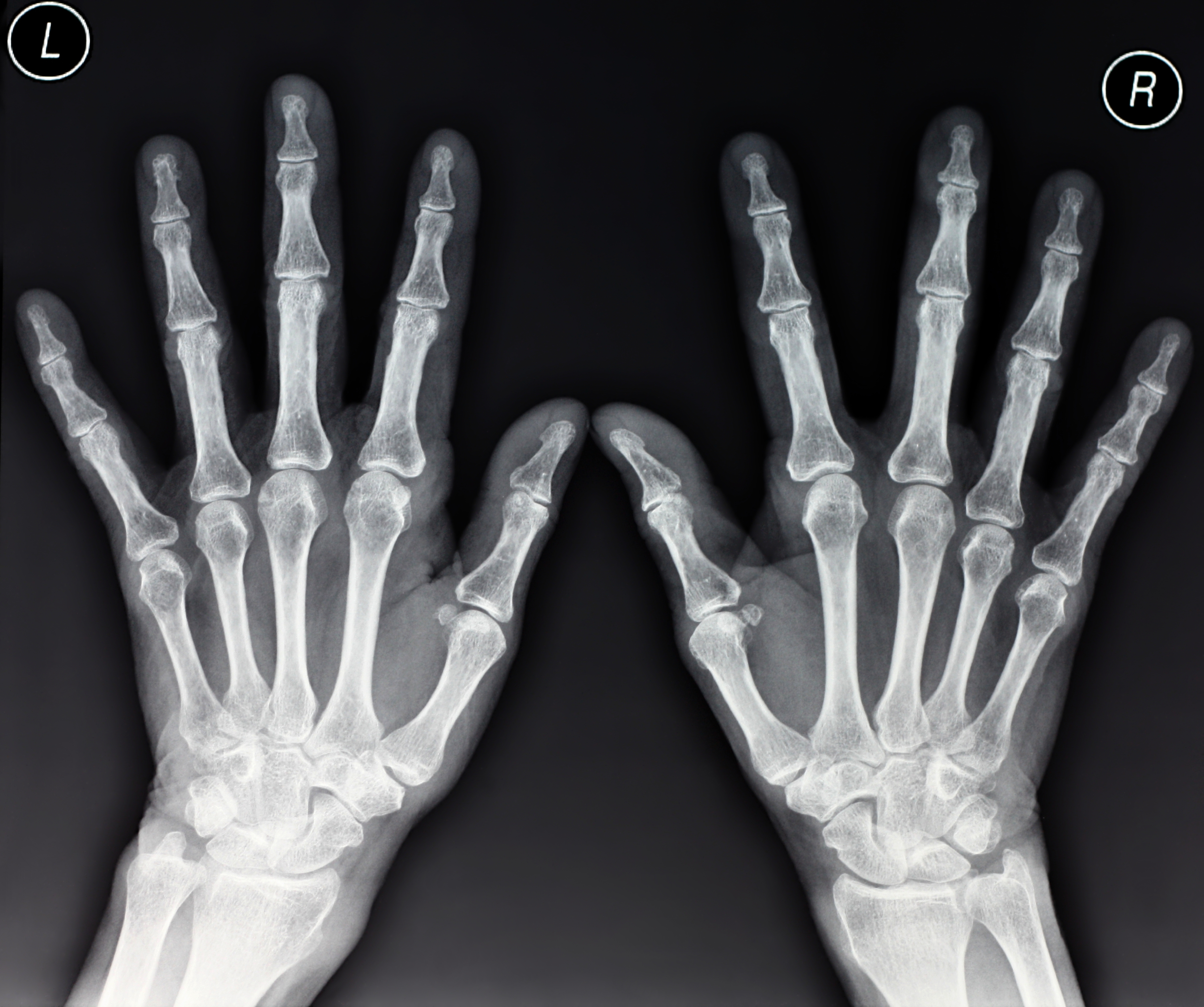
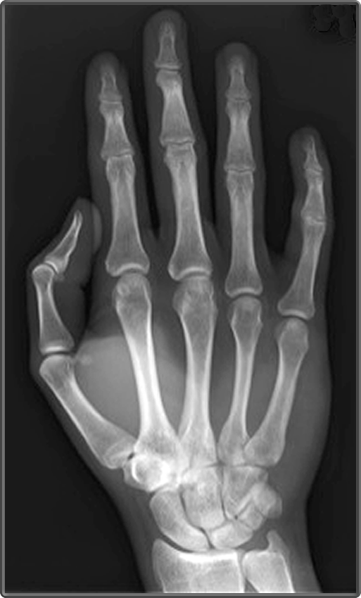
Symmetrical joints where the bones do not overlap except the carpal bones and the base of the metacarpal bones. Scaphoid fracture ulnar deviation view. The hand series consists of a posteroanterior oblique and a lateral projection.
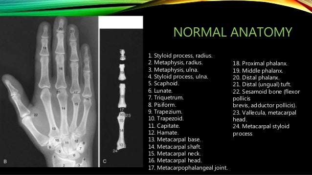
Adult hand radiographic anatomy. Anatomy function anatomy of the hand and wrist. Bones of the hand normal x ray pa finger bones articulate at the metacarpophalangeal joints mcpj the proximal interphalangeal joints pipj and the distal interphalangeal joints dipj the fingers each have 3 phalanges proximal middle and distal.
The joint spaces of the cmc joints are equal average 1 2 mm and form a zigzag configuration fig. Parts of the x ray tube. X rays show no bone.
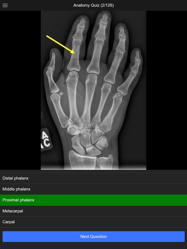
Purposegames lets you create and play games. In this case 2 extra views are added to the standard views oblique and pa with ulnar deviation. Diagnosis certain diagnosis certain.
See anatomy pictures of the 27 bones in the hand and wrist how they are connected with tendons and muscles and the nerves that run through the skeletal structure. Adjustments in kvp and mas should be considered in cases involving splints casts wraps swelling braces etc. Students teachers and rockstars alike all come here to create and learn.
Adult hand radiographic anatomy. The series primarily examines the radiocarpal and distal radioulnar joints the carpals metacarpals and phalanges. For each image you will have four possible answers to choose from.
This is a quiz called anatomy of an hand xray. Normal radiographic anatomy of the hand. X rays are indicated if there is post traumatic wrist pain with anatomical snuff box tenderness.
Bones muscles tendons nerves. Although x ray machines vary the general kvp ranges for radiography of the wrist and hand is between 50 65 kvp. Bones muscles tendons nerves.
Case contributed by dr benoudina samir. In this picture game you will label the structures of the upper limb from a series of x rays. Although additional radiographs can be taken for specific indications.
This is an interactive game to test your knowledge on the anatomy of the upper limb. Keep the body part as close to the cassette as possible in order to reduce oid object image distance. Stanford bone tumor bayesian network issssr msk lectures for residents ocad msk cases from around the world stanford msk mri atlas has served almost 800000 pages to users in over 100 countries.
Wrist Bone Anatomy X Ray Scan Stock Photos And Images
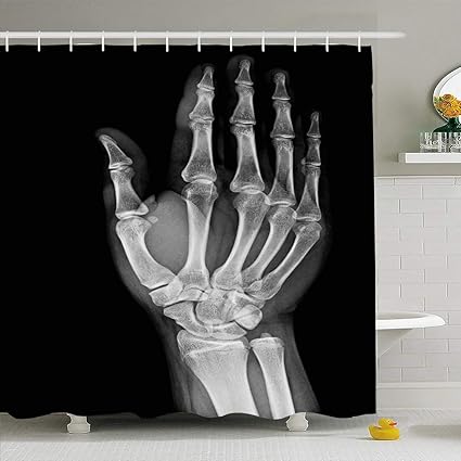 Amazon Com Ahawoso Shower Curtains 72 X78 Arm Xray Normal
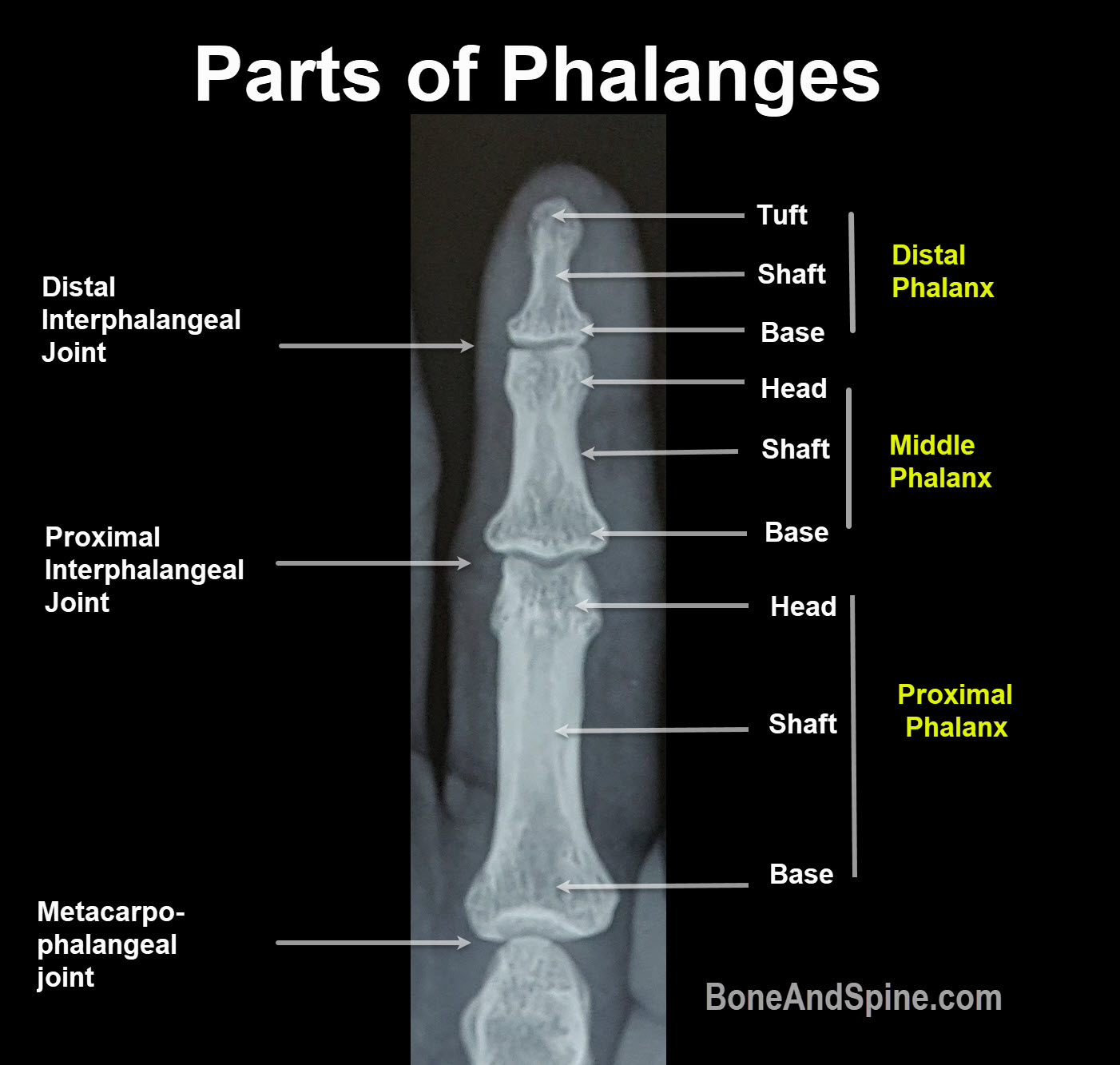 Phalanx Of Hand Anatomy And Function Bone And Spine
 Manus X Ray Anatomy Radiology Radiographic Stock Photo Edit
 Medical Imaging Technology Radiographic Anatomy Of Hand
 Hand Anesthesia Key
 Carpal Ossification Radiology Case Radiopaedia Org

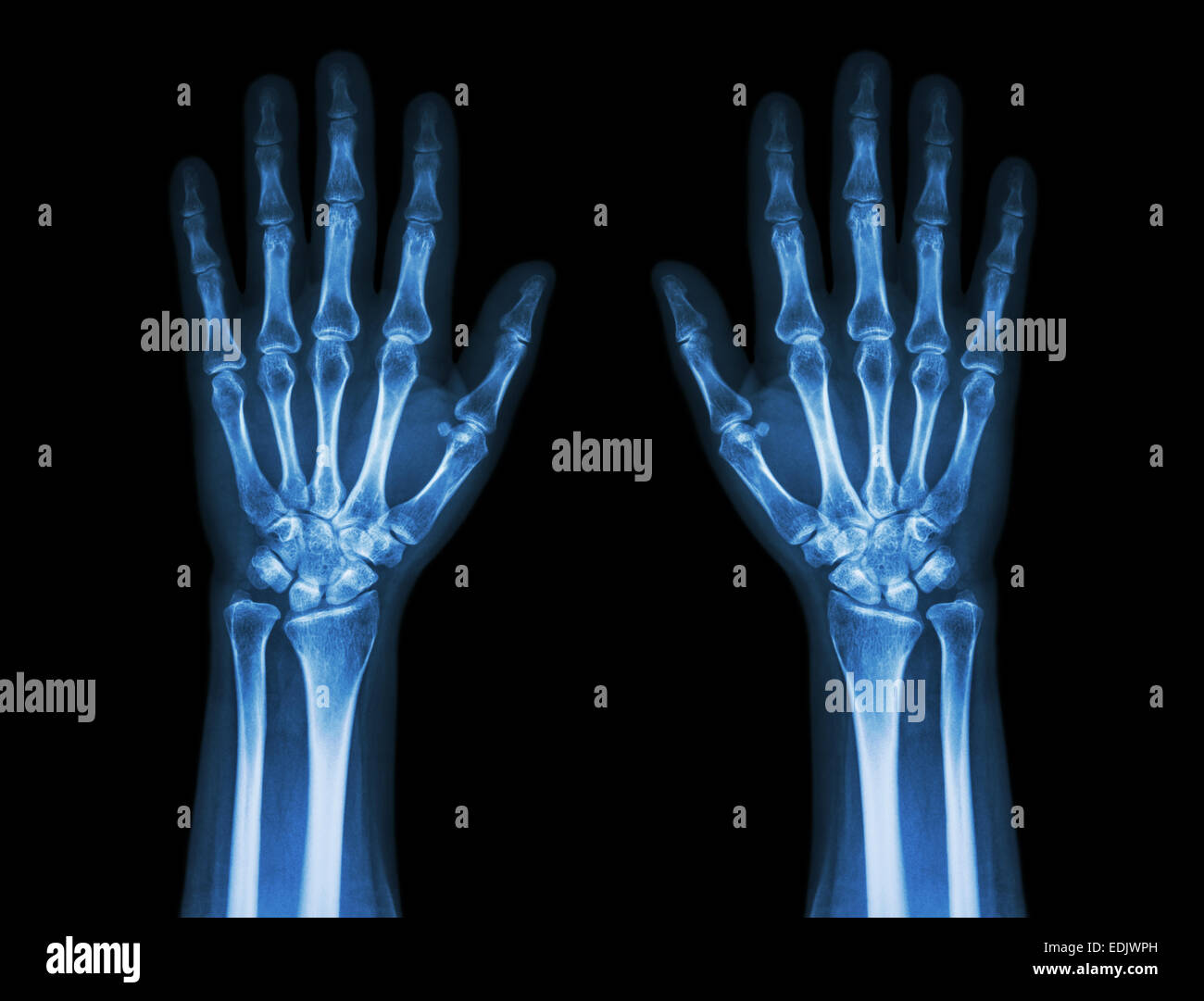 Human Wrist Anatomy Xray View Stock Photos Human Wrist
 X Ray Of Wrist And Hand
 Human Hand Bones X Ray Anatomy

 Ubc Radiology On The App Store
 X Ray Of Wrist And Hand
 Stock Illustration
 Anatomy Of An Hand Xray Purposegames

 The Importance Of Radiopaque Markers In Digital X Ray
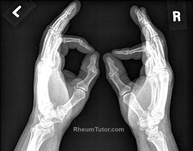 Approach To Hand X Rays Rheumtutor

 Approach To Hand X Rays Rheumtutor
 Foot X Rays
Belum ada Komentar untuk "X Ray Hand Anatomy"
Posting Komentar