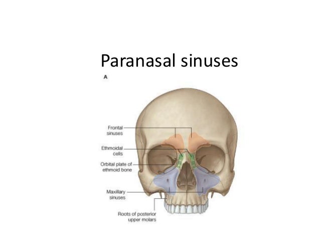
Paranasal Sinus Anatomy
Posterior 13rd is bony 1. The maxillary sinuses the largest of the paranasal sinuses are under the eyes.
![]() Paranasal Sinuses Anatomy And Clinical Aspects Kenhub
Paranasal Sinuses Anatomy And Clinical Aspects Kenhub
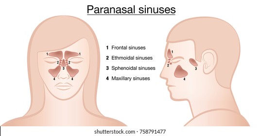
Humans possess four paired paranasal sinuses divided into subgroups that are named according to the bones within which the sinuses lie.
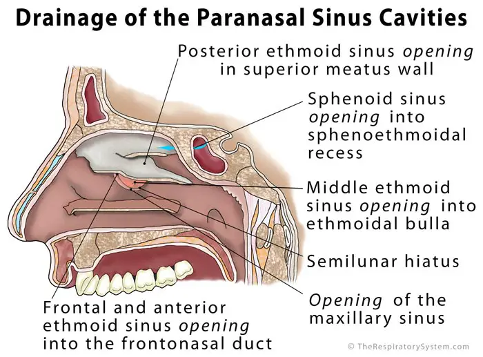
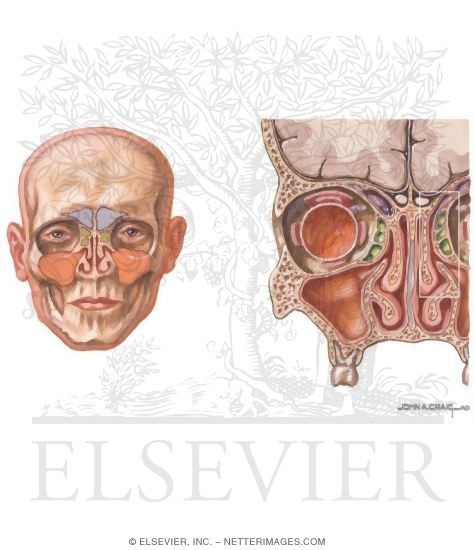
Paranasal sinus anatomy. External nose it is pyramidal in shape with its root up and the base directed downwards nares external opening of nose choanae open into the nasopharynx 1. Infection of the sinuses causes inflammation particularly pain and swelling of the mucosa and is known as sinusitis. Anatomy of the equine paranasal sinuses.
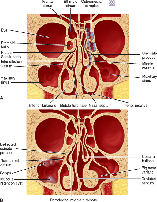
The nasal septum separates the left and right maxillary sinuses 9. Ethmoid air cells sinuses. Paranasal sinuses frontal sinuses.
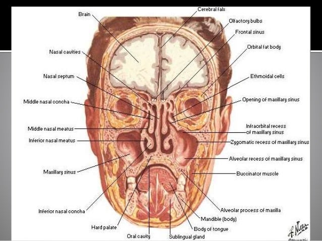
From the nasal mucosa and their continued communication with the nasal fossae. The superior border of this sinus is the bony orbit the inferior is the maxillary alveolar bone and corresponding tooth roots. Others are much smaller.
If more than one sinus is affected it is called pansinusitis. Protection of internal cranial structures maintaining structural strength without a significant increase in weight augmenting vocalisation. The sinuses are a connected system of hollow cavities in the skull.
Your cheekbones hold your maxillary sinuses the. The paranasal sinuses of mammals form complex air filled structures and probably exist for a variety of functions including but not exhaustively. The maxillary sinuses are the largest of the all the paranasal sinuses.
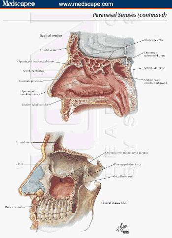
The largest sinus cavities are about an inch across. Where are the paranasal sinuses situated. The most posterior of all the paranasal sinuses these are located behind the ethmoid sinuses near the pituitary gland and optic nerves 8.
The frontal sinuses superior to the eyes in the frontal bone. In the frontal ethmoid and sphenoid of the cranium and the maxillary bones of the face. As the paranasal sinuses are continuous with the nasal cavity an upper respiratory tract infection can spread to the sinuses.
What are the paranasal sinuses a formation from. They have thin walls which are often penetrated by the long roots of the posterior maxillary teeth. Sphenoid sinus within the sphenoid bones.
External nose internal nose musculature of nose vascular supply nervous innervation and lymphatic drainage paranasal sinuses 3. Histology of the nose and paranasal sinuses. The nasal cavity and all paranasal sinuses are lined by respiratory pseudostratified epithelium except areas of the roof of the cavity most superior part of nasal septum and the medial part of superior turbinate are lined by mucosa that contains receptors for the smell sensation known as olfactory area.
 Paranasal Sinuses Images Stock Photos Vectors Shutterstock
 38 Maxillary Sinus Anatomy Pathology And Graft Surgery
 Ct Anatomy Of Para Nasal Sinuses
 Paranasal Sinus Cavities Acland S Video Atlas Of Human Anatomy
![]() Paranasal Sinuses Anatomy And Clinical Aspects Kenhub
Paranasal Sinuses Anatomy And Clinical Aspects Kenhub
 Sinuses Of Nose
 Nose And Sinus Anatomy Thomas S Higgins Md Msph
 Paranasal Sinus An Overview Sciencedirect Topics
 Surgical Views Paranasal Sinus Disease In Horses Ce
 Sinusitis Cancer Therapy Advisor
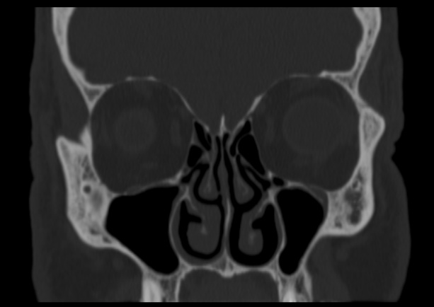
 A Ct Scan In Coronal Plane Shows The Normal Paranasal Sinus
 Paranasal Sinus Anatomy What The Surgeon Needs To Know
 Paranasal Sinuses
 Paranasal Sinuses
 Beautifully Designed Paranasal Sinus Art Fine Art America
 The Hidden Anatomy Of Paranasal Sinuses Reveals
 Paranasal Sinuses Radiology Key
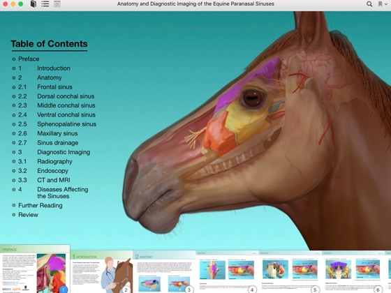 Anatomy And Diagnostic Imaging Of The Equine Paranasal
Paranasal Sinus Definition Location Anatomy Function
 The Paranasal Sinuses Structure Function Teachmeanatomy

 Basic Ct Anatomy Of Paranasal Sinuses
 Startradiology
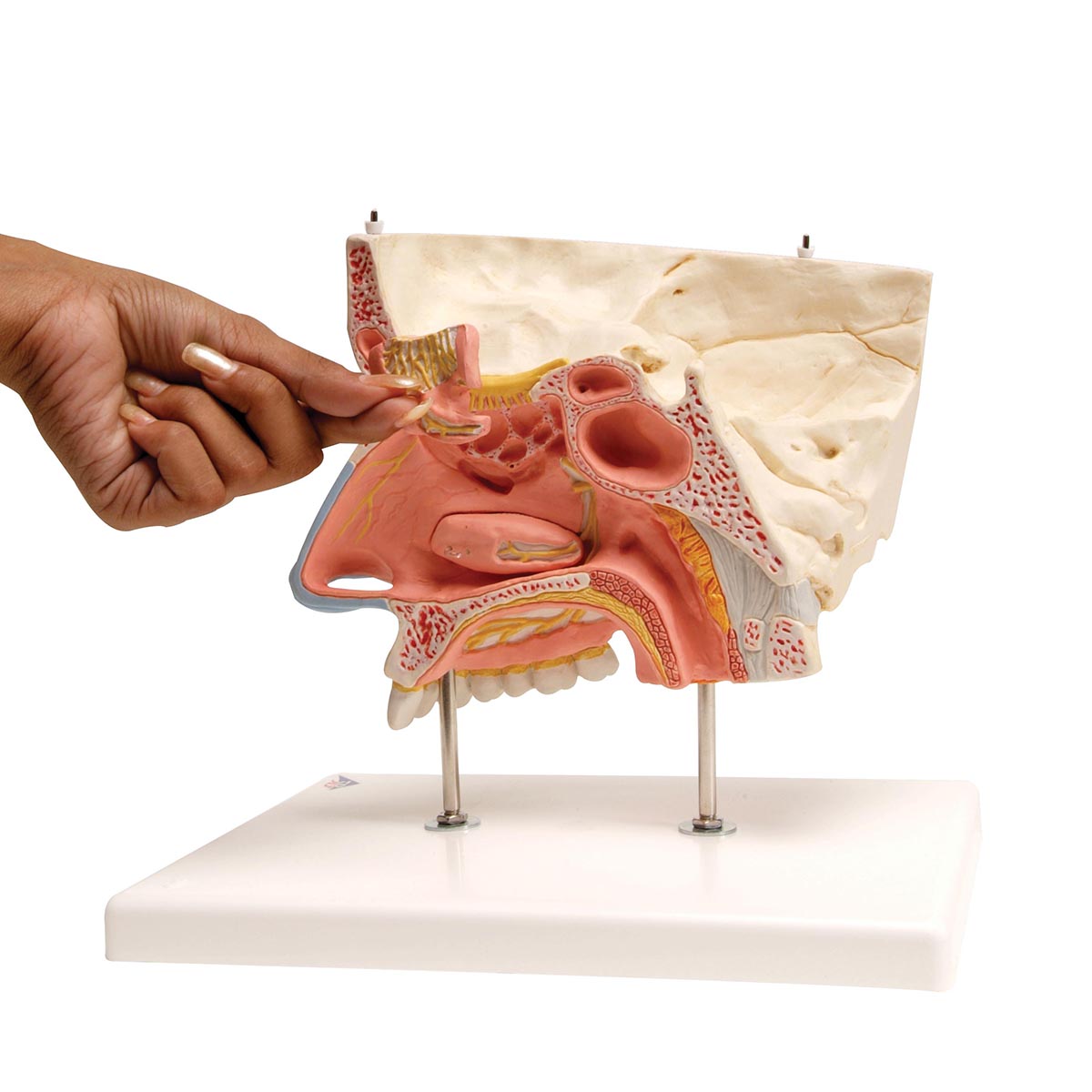 Human Nose Model With Paranasal Sinuses 5 Part 3b Smart
 Overview And Topographic Anatomy Of The Paranasal Sinuses
 Paranasal Sinuses
 A Practical Approach To The Patient With Sinusitis
 Paranasal Sinus Anatomy
Belum ada Komentar untuk "Paranasal Sinus Anatomy"
Posting Komentar