Macula Anatomy
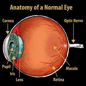
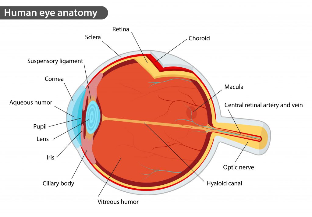
The macula is a circular area of diameter 55 mm with a center located 17 degrees or 40 50 mm temporal and 053 08mm inferior to the center of the optic disc. Anatomy of macula 1.
 Eye Anatomy
The macula is a small but important area in the center of the retina.
/macular_degeneration-56a9cf7f3df78cf772aab31f.jpg)
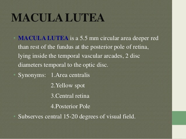
Macula anatomy. Anatomy of internal ear in senses. Macula lutea macula lutea is a 55 mm circular area deeper red than rest. The bundle of nerve fibers at the back of the eye that carry visual messages from the retina to the brain.
When the gaze is fixed on any object the centre of the macula the centre of the lens and the object are in a straight line. The fovea is the area of best visual acuity. It is one hundred times more sensitive to detail than the peripheral retina.
Vestibular structures of sensory cells called a macula. It is marked by the presence of multiple cyst like cystoid formations which cause edema swelling in the macular area resulting in blurred or decreased central vision. Photopic color vision are primary functions of this area.
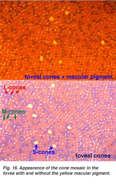
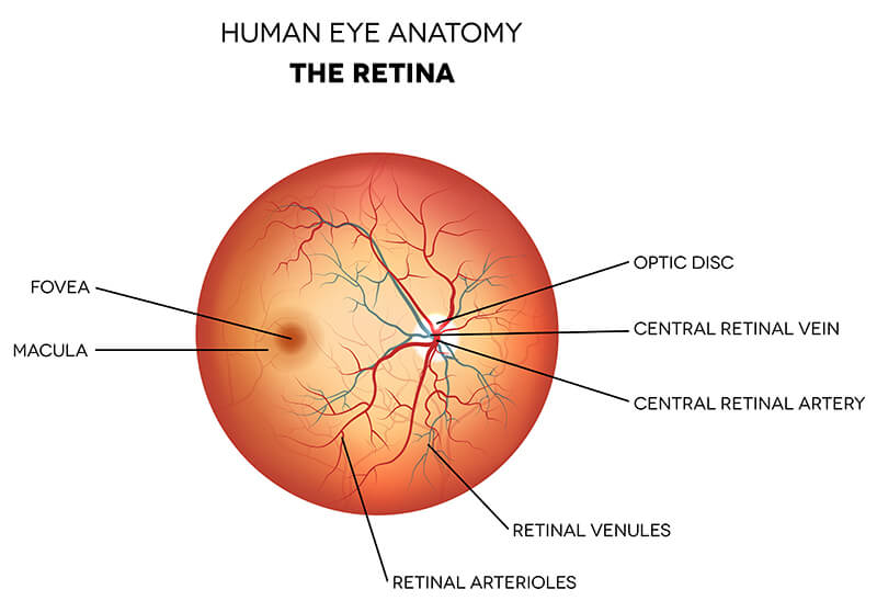
The light sensing nerve cells rods and cones located in the retina. It contains a large amount of conesnerve cells that are photoreceptors with high acuity. The normal central retinal artery black arrow is located nasal to the central retinal vein green arrow in the optic disc.
Mechanical senses of hair cells known as maculae. An area of the eye near the center of the retina where visual perception is most acute. The macula is the pigmented part of the retina located in the very center of the retina.
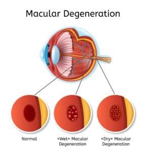
The macula or macula lutea is an oval shaped pigmented area near the center of the retina of the human eye and some other animalian eyes. Cystoid macular edema cme is a painless disorder affecting the macula. You need the macula to clearly see details of objects in front of you like faces and written text.
The macula in humans has a diameter of around 55 mm 022 in and is subdivided into the umbo foveola foveal avascular zone fovea parafovea and perifovea areas. Macula lutea in anatomy the small yellowish area of the retina near the optic disk that provides central vision. The macula is responsible for the sharp straight ahead vision that is used for seeing fine detail reading driving and recognizing faces.
Contents anatomy of macula lutea embryology blood supply macular function tests. The portion of the eye at the center of the retina that processes sharp clear straight ahead vision. In the center of the macula is the fovea perhaps the most important part of the eye.
A number of eye problems can affect the macula and can lead to vision loss if they are not treated. Vestibule of sensory cells called a macula which is about 2 mm 008 inch in diameter.
 Anatomy Of The Eye 101 Eyecheck
 Simple Anatomy Of The Retina By Helga Kolb Webvision
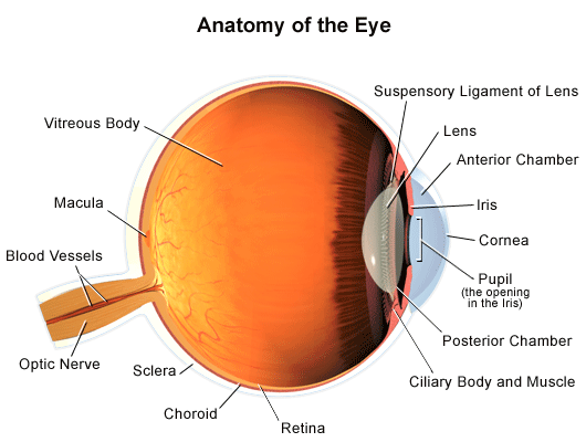 Anatomy Of The Eye
 Human Eye Macula Of Retina Conjunctiva Anatomy Png Clipart
 Macular Degeneration What Is It And How Can It Be Treated
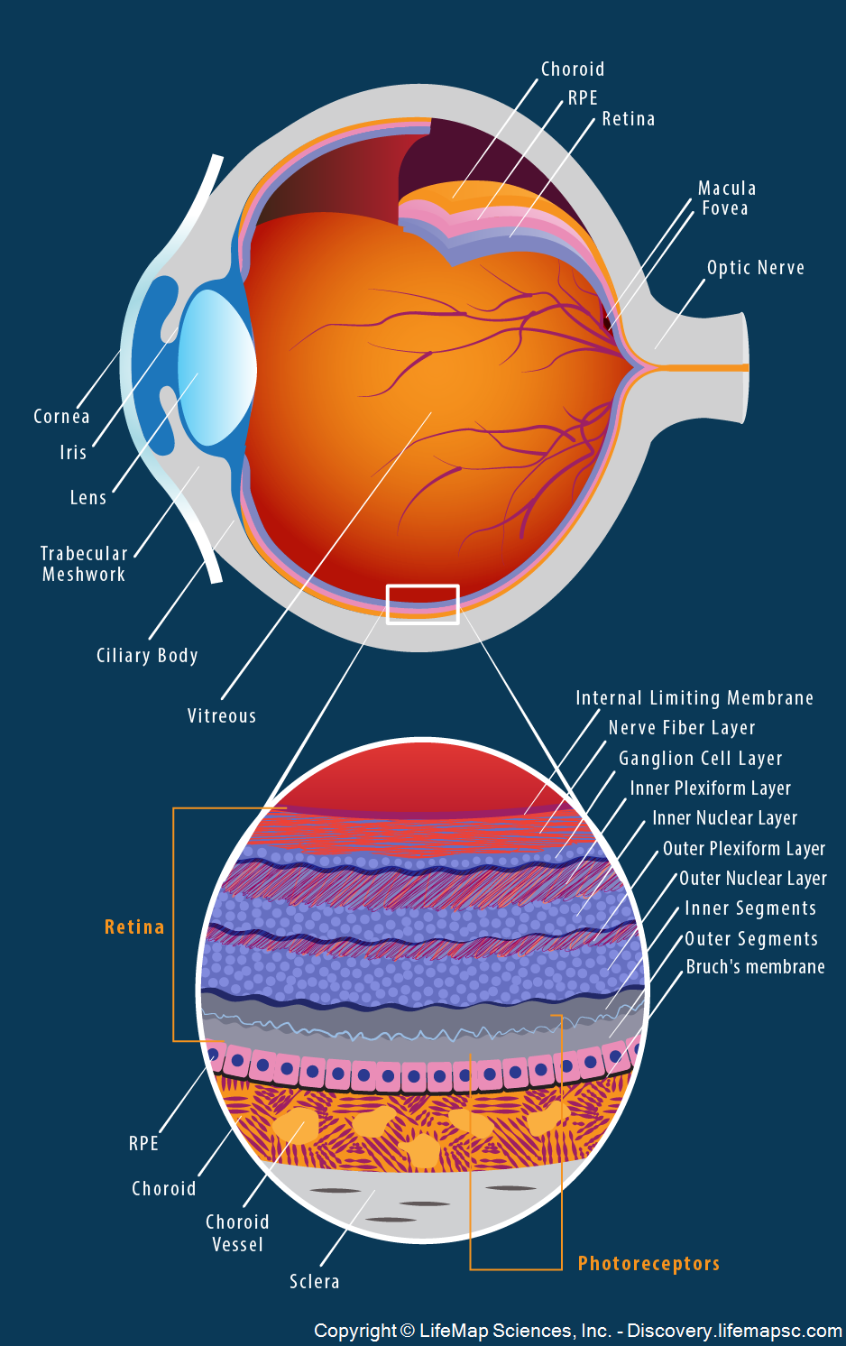 Human Eye Anatomy Infographic Lifemap Discovery
 What Is The Macula What The Macula Does
Macula Anatomy Function And Significance
 Eye Anatomy How Youniggasook Od Ciliary Body Retina Iris
 Eye Anatomy And How The Eye Works
:max_bytes(150000):strip_icc()/GettyImages-479379785-e7c7c41ed3574869879c1ca6ef2defa3.jpg) Macular Telangiectasia Types And Symptoms
Macular Telangiectasia Types And Symptoms
 Anatomy Of Macula
 How The Eye Works As A Camera Amdf
 Simple Anatomy Of The Retina By Helga Kolb Webvision
 Eye Anatomy Neurology Medbullets Step 1
 Fovea Art Print Eye Anatomy Poster Macula Lutea Histology Watercolor Optometry Illustration Ophthalmology Art Fovea Showing Muller Cell Cone
 About Basic Eye Anatomy Gem Clinic Glaucoma Eye
 Retina Farmington Retina Specialist Ct Consulting
 Eye Exercises For Best Vision
 Lasik
Lasik
 Retina Diseases Milwaukee Macular Degeneration Mequon
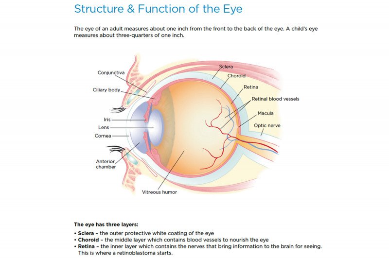 Retinoblastoma Anatomy Of The Eye Memorial Sloan
 Colorado Retina Patient Education
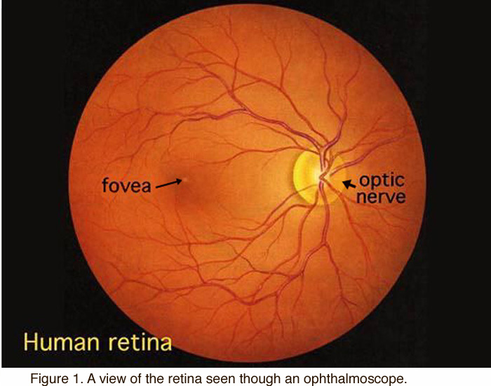
 Normal Retinal Anatomy The Retina Reference
 Macula American Academy Of Ophthalmology
 Retina Macula Lutea Choroid Anatomy Eyeball Anatomy Eye

Belum ada Komentar untuk "Macula Anatomy"
Posting Komentar