Iliac Anatomy
It divides into two branches. The iliac crest bone marrow and bone grafts.
 Ovid Lippincott Williams Wilkins Atlas Of Anatomy Psoas
The common iliac vein created by the union of the internal and external iliac veins forms in the abdomen at the level of the fifth lumbar vertebrae.
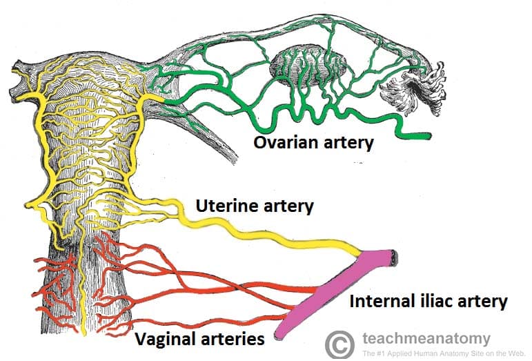
Iliac anatomy. The aorta ends at the fourth vertebra of the lumbar spine. The femoral vein is renamed the external iliac vein once it passes the inguinal ligament. The internal iliac vein supplies blood to the visceral organs in the pelvic region.
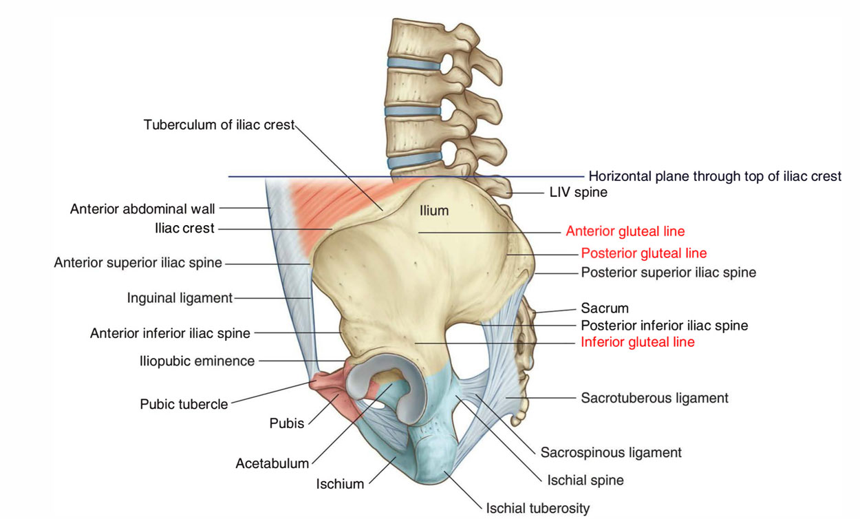
It is located on the superior and lateral edge of the ilium very close to the surface of the skin in the hip region. The vesicular branches of the internal iliac arteries supply the bladder it is a short thick vessel smaller than the external iliac artery and about 3 to 4 cm in length. Hip pointer injury to the iliac crest.
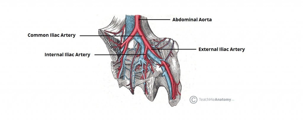
Athletes may only become familiar with. The common iliac artery originates from the abdominal aorta the main blood vessel in the abdominal area. The blood supply to the body arises from the aorta the main outflow of the left ventricle of the heart.
The iliac recess is a concavity dorsolateral to the sharply curving posterior end of the internal ilio ischiatic crest. The external iliac connects to the femoral veins. The iliac crest has a large amount of red bone marrow and thus it is the site of bone marrow harvests from both sides to collect the stem cells used in bone marrow transplantation.
Bruce holmes seat of wound abdominal wall a highest point of iliac crest. Both the aorta and the systemic arteries are part of the systemic circulatory system which carries oxygenated blood from the heart to the other areas of the body and back. The internal iliac artery supplies the walls and viscera of the pelvis the buttock the reproductive organs and the medial compartment of the thigh.
Summary the common iliac vein is formed by the unification of the external and internal iliac vein. The iliac crest is the curved superior border of the ilium the largest of the three bones that merge to form the os coxa or hip bone. Hip pads to prevent injury to the iliac crest.
Variation in the muscles and nerves of the leg in e. Anatomy of the iliac crest synonyms. The abdominal aorta gives rise to two common iliac arteries at its termination which then further divided into the external and internal iliac arteries.
The two common iliac veins unite to form the inferior vena cava. The internal iliac arteries are the major arteries of the pelvis and together with their many branches supply the blood to the major organs and muscles of the pelvis. The iliac crest is also considered the most ideal donor site for bone grafting when a large quantity of bone is needed.
The internal iliac vein drains the. The iliac crest is the upper border of the wing of the ilium. The internal iliac arteries are branches of the common iliac arteries which themselves are branches from the aorta.
:background_color(FFFFFF):format(jpeg)/images/library/12623/Common_iliac.png) Iliac Arteries Branches And Clinical Points Kenhub
Iliac Arteries Branches And Clinical Points Kenhub
 Iliac Crest Fracture Apophysis Graft Pain Causes
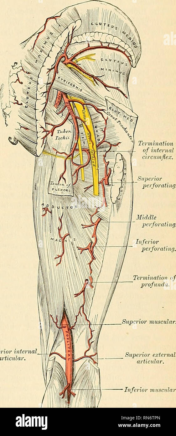 Anatomy Descriptive And Applied Anatomy The Internal
 Arteries Of The Pelvis Internal Iliac Pudendal Vesical
Surgical Management Of Intractable Pelvic Hemorrhage Glowm
:watermark(/images/watermark_only.png,0,0,0):watermark(/images/logo_url.png,-10,-10,0):format(jpeg)/images/anatomy_term/common-iliac-artery/3j4tc88p2G8vUomS70mw_A._iliaca_communis_02.png) Iliac Arteries Branches And Clinical Points Kenhub
Iliac Arteries Branches And Clinical Points Kenhub
Anatomy Stock Images Torso Musculus Quadratus Lumborum
 Iliac Crest Images Stock Photos Vectors Shutterstock
 The Muscles And Fasciae Of The Iliac Region Human Anatomy
 Superficial Circumflex Iliac Artery Wikipedia
 Iliac Artery Anatomy Britannica
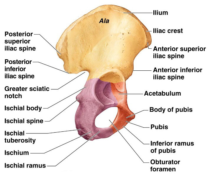 Hip Bone Anatomy Or Pelvic Bone Ilium Pubis Ischium Bone
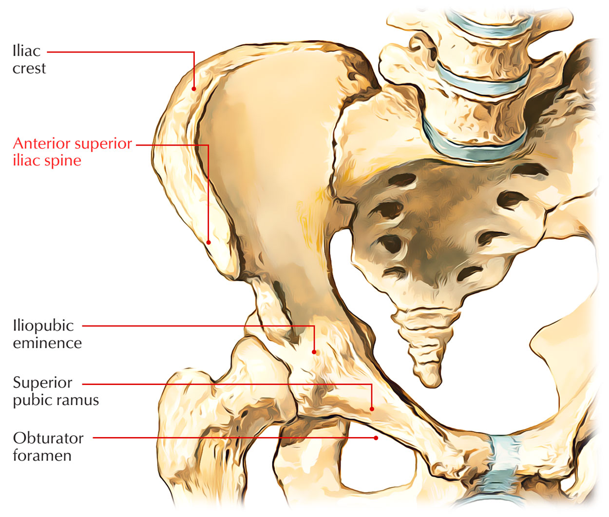 Anterior Superior Iliac Spine Earth S Lab
 Thigh Blood Supply Anatomy Medbullets Step 1
 Intraperitoneal And Retroperitoneal Anatomy The 3rd
 25 Best Memes About Posterior Superior Iliac Spine
 Arteries Of The Pelvis Internal Iliac Pudendal Vesical

 Reconstruction Reduction Fixation Ao Surgery Reference
The Anatomy Of The Laboratory Mouse

Belum ada Komentar untuk "Iliac Anatomy"
Posting Komentar