Connective Tissue Anatomy
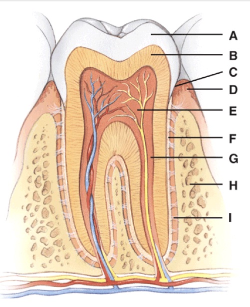
These canals consist of blood vessels that branch off the central vessels. Human anatomy and physiology tissues.
 E Book 03 Connective Tissue Supports And Protects
Sparsely distributed chondrocytes in matrix.

Connective tissue anatomy. Branching off the central canal at right angles are perforating canals. Matrix is dominated by chondroitin sulfate and collagen. Found in the upper portion of the respiratory tract trachea and bronci ends of bones and ribs skeleton of a fetus.
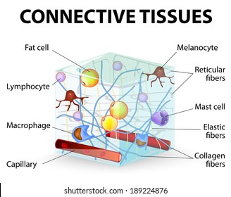
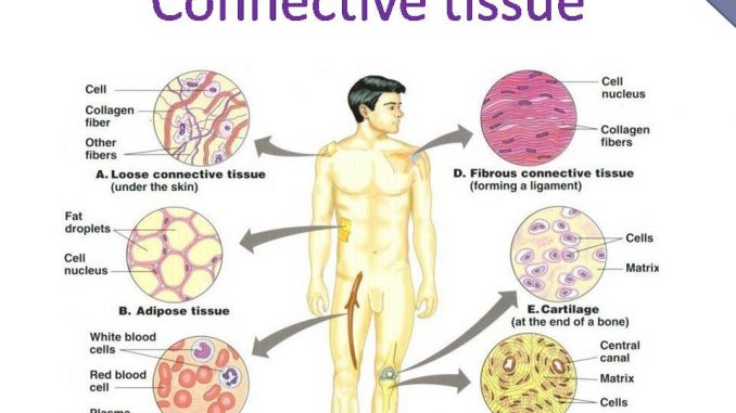
Adipocytes account for most of volume. Although it is the most abundant and widely distributed of the primary tissues the amount of connective tissue in a particular organ varies. Loose connective tissue is named so because of the weave and type of its constituent fibers.
These fibers form an irregular network with spaces between the fibers. Nearly all the fat in the body. Highly vascularized fat that breaks down to provide heat to the blood.
Four types of connective tissue 2342 all develop from mesenchyme 3295 different degrees of blood flow 3457 extracellular matrix full of ground substance and fibers 3594. Regular irregular and elastic. Three fibers in connective tissue.
Eachosteon consists of a central canal which contains blood vessels and nerves surrounded by concentric rings lamellae of hard matrix and collagen fibers. Connective tissue ct is a one of the four main classes of tissues. Produces both the fibers and the ground substance of the extracellular matrix.
Skeletal voluntary muscles smooth muscles and the cardiac muscle in the heart. Connective tissue binds the cells and organs of the body together and performs many functions especially in the protection support and integration of the body. Collagen resist tension thick has 3 strands reticular provides structural support thin elastic enables recoil of stretched tissues.
Muscle tissue which responds to stimulation and contracts to provide movement is divided into three major types. It holds organs in place and attaches epithelial tissue to other underlying tissues. A type of loose connective tissue proper.
Ligaments and tendons are made of dense regular connective tissue. Most abundant type of cartilage. Dense regular connective tissue fibers are parallel to each other enhancing tensile strength and resistance to stretching in the direction of the fiber orientations.
There are three major categories of dense connective tissue. Like to the timber framing of a house the connective tissue provides structure and support throughout the body. In vertebrates the most common type of connective tissue is loose connective tissue.
 Connective Tissue Boundless Anatomy And Physiology
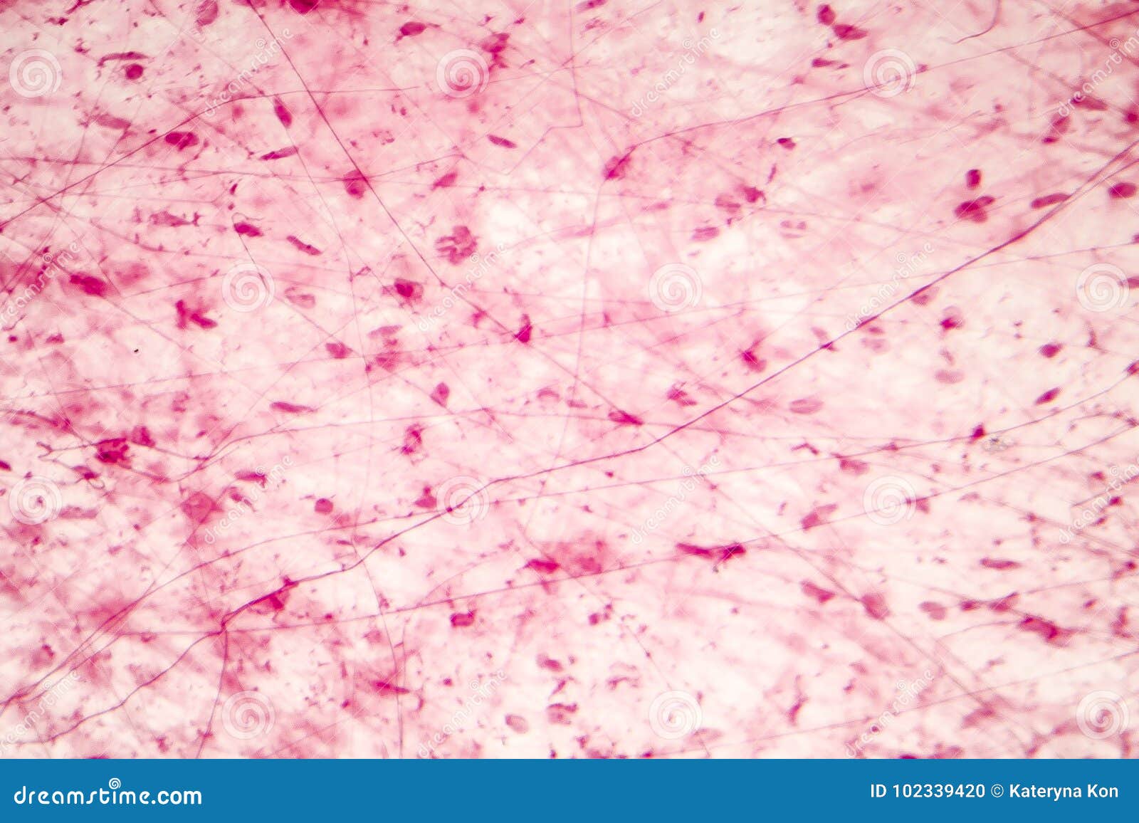 Loose Connective Tissue Stock Photo Image Of Anatomy
 Connective Tissue Images Stock Photos Vectors Shutterstock
 Connective Tissues Course Hero
 Skeletal Muscle Physiology
 Anatomy And Cell Biology 3309 Lecture Notes Summer 2017
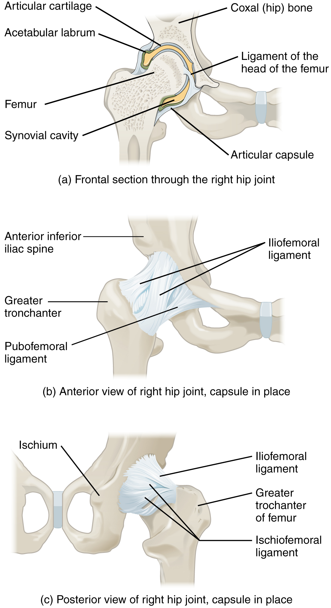 Joints Ligaments And Connective Tissues Advanced Anatomy
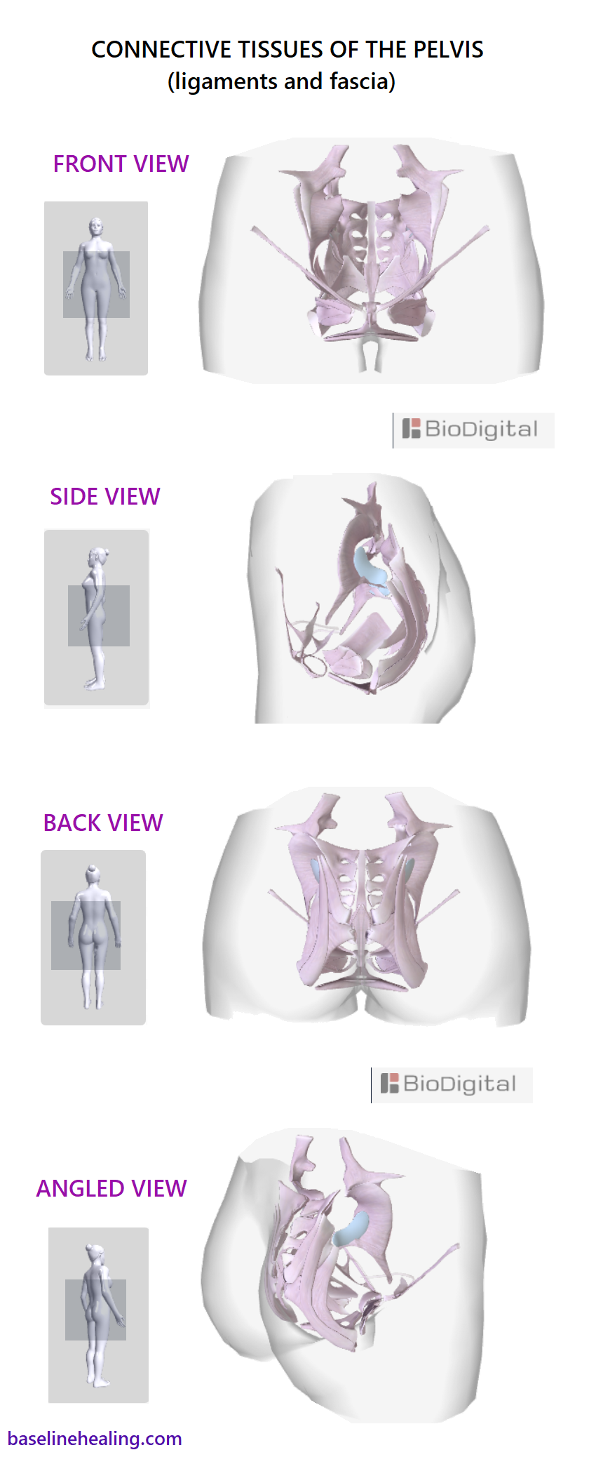 The Pelvis Anatomy Images Pelvic Floor Connective Tissues
 Ch04 Mineralized Connective Tissues
 Animal Primary Tissues Boundless Biology
 Muscle Tissue Anatomy Physiology Nutrition With External
 Connective Tissue Anatomy Physiology 121 With Mazin At
 Loose Areolar Connective Tissue Areolar Ct Loose Ct Fibers
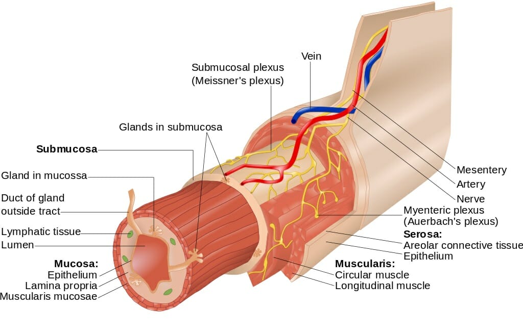 Lamina Propria Definition Function And Structure Biology
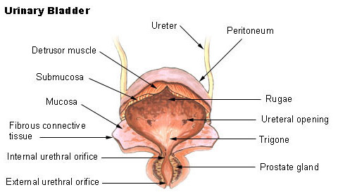 Seer Training Urinary Bladder
 Description
 Anatomy And Physiology Connective Tissue Notes
 Types Of Tissues Anatomy And Physiology I
 Connective Tissues Useful Notes On The Connective Tissues
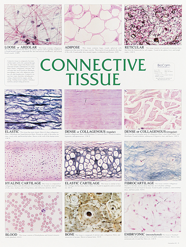 Connective Tissue Chart
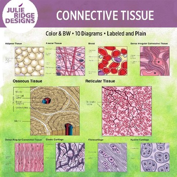 Human Anatomy Connective Tissue Diagrams By Julie Ridge
 Connective Tissue Britannica
 This Consists Of Calcified Connective Tissue Proprofs
 Histology Flashcards Connective Tissue Learn All Organs
 Connective Tissue Characteristics Functions And Types

Belum ada Komentar untuk "Connective Tissue Anatomy"
Posting Komentar