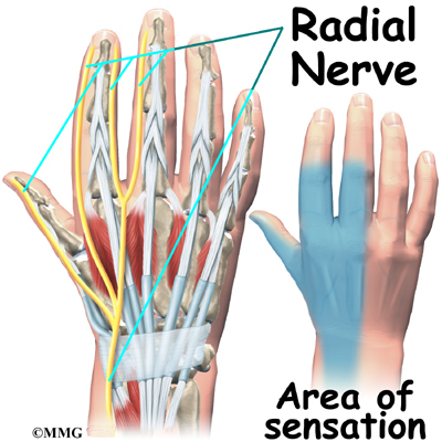
Anatomy Of The Wrist
It is actually a collection of multiple bones and joints. A patients guide to wrist anatomy introduction.
 Carpal Tunnel Syndrome Symptoms And Treatment Orthoinfo
The mass that results from these bones is called the carpus.

Anatomy of the wrist. Superficialis tendons which pass through the palm side of the wrist and hand. The wrist is the junction of the distal end of the radiusulna and the adjacent carpal bones. The anatomy of the wrist joint is extremely complex probably the most complex.
The important structures of the wrist can be divided into several categories. The carpus is rounded on its proximal end where it articulates with the ulna and radius at the wrist. The bones comprising the wrist include the distal ends of the radius and.
The wrist is a complex joint that bridges the hand to the forearm. There are eight small carpal bones in the wrist that are firmly bound in two rows of four bones each. The wrist connects the hand to the forearm.
The carpal bones are arranged in 2 interrelated rows. It is often compared to the ankle joints in structure however through evolution the wrist has become more delicate and lost many of the characteristics that would allow it to be a truly effective weight bearing joint. The main tendons of the hand are.
The wrist is a complex system of many small bones known as the carpal bones and ligaments. One row connects with the ends of the bones in the forearmthe radius and ulna. Extensor tendons of the fingers which attach to the middle and distal phalanges and extend.
There are two long bones in the forearm that run from the elbow to the wrist. As you can see the wrist is a complex area of the body. Osteology of the wrist.
The smaller bone the ulna is on the little finger side. It consists of the distal ends of the radius and ulna bones eight carpal bones and the proximal ends of five metacarpal bones. In human anatomy the wrist is variously defined as 1 the carpus or carpal bones the complex of eight bones forming the proximal skeletal segment of the hand.
The wrist joint also known as the radiocarpal joint is a synovial joint in the upper limb marking the area of transition between the forearm and the hand. The larger bone the radius is on the same side as the thumb. 2 the wrist joint or radiocarpal joint the joint between the radius and the carpus and 3 the anatomical region surrounding the carpus including the distal parts of the bones of the forearm and the proximal parts of the metacarpus or five metacarpal bones and the series of joints between these bones thus referred to as wrist joints.
Profundus tendons which pass through the palm side of the wrist and hand.
 Anatomy Of Hand Wrist Bones Muscles Tendons Nerves
 Wrist Anatomy Bones Ligaments Muscles Nerves
 Common Hand And Wrist Conditions Pro Sports Orthopedics
 Wrist Joint Anatomy Overview Gross Anatomy Natural Variants
 Hand And Wrist Anatomy Baxter Regional Medical Center
 Wrist Anatomy Wrist Bones And Movements Kenhub
 Carpal Tunnel Syndrome Cleveland Clinic
 Anatomy Of The Wrist Thumb And Hand Musculoskeletal Key
 Wrist Joint Anatomy Overview Gross Anatomy Natural Variants
 Wrist Anatomy Orthopedic Surgery Algonquin Il
 Wrist Anatomy Images Stock Photos Vectors Shutterstock
 Hand Wrist Anatomy Lakeshore Orthopaedics
 Wrist Bones Kirkland Wa Evergreenhealth
 1 Topographic Anatomy Of The Wrist A Dorsal Surface Of
 Wrist Anatomy Images Stock Photos Vectors Shutterstock
 Understanding The Anatomy Of The Wrist Bodyheal
 Hand And Wrist Anatomy Murdoch Orthopaedic Clinic
 The Forearm Wrist And Hand Musculoskeletal Key
 The Anatomy Of Wrist Related Muscles A Anterior View B
 Nerves Blood Vessels And Lymph Advanced Anatomy 2nd Ed
 Wrist Muscular Anatomy Musculoskeletal Learning Portfolio
 Hand Anatomy Midwest Bone Joint Institute Elgin Illinois


Belum ada Komentar untuk "Anatomy Of The Wrist"
Posting Komentar