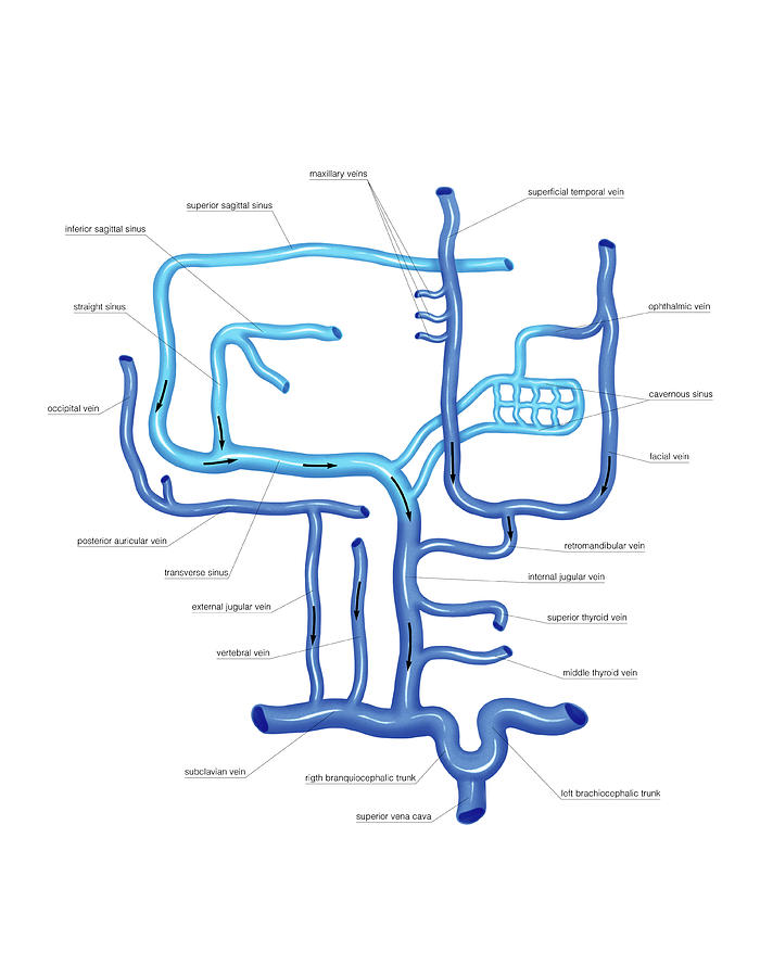
Anatomy Of Venous System
The physiology of venous return. We distinguish between the superficial and the deep venous systems.
The arteries divide into smaller vessels called arterioles.

Anatomy of venous system. Both the fluid components and the vessels through which they flow reach their greatest elaboration and specialization in the mammalian systems and particularly in the human body. As arterial blood flows into the leg distal superficial veins constantly fill venous blood is regularly emptied from the superficial system into the deep venous system via the sfj spj and perforators this blood is then returned to the right side of the heart through one way valves by calf muscle contraction. This system contains two fluids blood and lymph and functions by means of two interacting modes of circulation the cardiovascular system and the lymphatic system.
They carry the blood at high pressure away from the heart. Blood is the transport media of nearly everything within the body. Anatomy of the venous system the venous system is that part of the circulation in which the blood is transported from the periphery back to the heart.
The soleal veins are deep venous sinuses and veins of the soleus muscle that drain into the popliteal vein. Your venous system is a network of veins that carry blood back to your heart from other organs. Veins carry deoxygenated blood to the lungs where they receive oxygen.
They are an important part of the calf muscle pump mechanism see ch. The circulatory and lymphatic systems are networks of vessels and a pump that transport blood and lymph respectively throughout the body. It transports hormones nutrients oxygen antibodies and other important things needed to keep the body healthy.
The anatomy and physiology of venous return are described with an emphasis on the differences between standing and walking and the interplay between the venous systems of both the foot and the calf. Aorta is the largest artery. When these systems are infected with a microorganism the network of vessels can facilitate the rapid dissemination of the microorganism to other regions of the body sometimes with serious results.
Arteries are the strongest thickest and most elastic of all the vessels. The paired veins join into common trunks in the upper calf before forming the below knee popliteal vein.
 Medical Description Of The Venous System Of Blood Stock
 Lower Extremity Venous Reflux Baliyan Cardiovascular
 Venous Drainage Anatomy Overview Microscopic Anatomy
 Transmedullary Venous Anastomoses Anatomy And Angiographic
 Venous Drainage Of The Brain Anatomy Geeky Medics
 Venous System Of The Upper Limb
 Science Source Venous System Of The Trunk Artwork
 The Venous System Ppt Download
 Misplaced Central Venous Catheters Applied Anatomy And
 Venous System Of The Upper Limb Artwork Stock Image
 Human Anatomy Art Print Venous System Artery Vintage Anatomy Doctor Medical Art Antique Book Plate T Shirt By Frenchfineart
 Biorender Venous System Male On Body
 Venous System Of The Head And Neck
 Anatomy Of The Portal Venous System Omitted Is The Anatomy
 Illustration Human Figure With Venous System Heart Vein
 Blood Supply And Venous Drainage Enlarged Thyroid
 Anatomical Structure Of Human Body Presented Detail Of Circulatory
 Blood Supply And Venous Drainage Of The Kidney Harold E
 Cardiovascular System Human Veins Arteries Heart
 Science Source Venous System Of The Chest Artwork
 Anatomy Of Lower Extremity Venous System In Khar West
 Solved 12 The Human Arterial And Venous Systems Are Diag
 Venous Insufficiency Redbacteria
 Anatomy Venous System Of The Heart
 Basic Anatomy Of The Venous System Of The Lower Extremities
 Venous Anatomy Of The Abdomen And Pelvis Clinical Gate
 Chapter 126 Development Of The Venous System The Inferior
 The Venous Drainage Of The Abdomen And Chest Flashcards


Belum ada Komentar untuk "Anatomy Of Venous System"
Posting Komentar