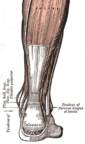
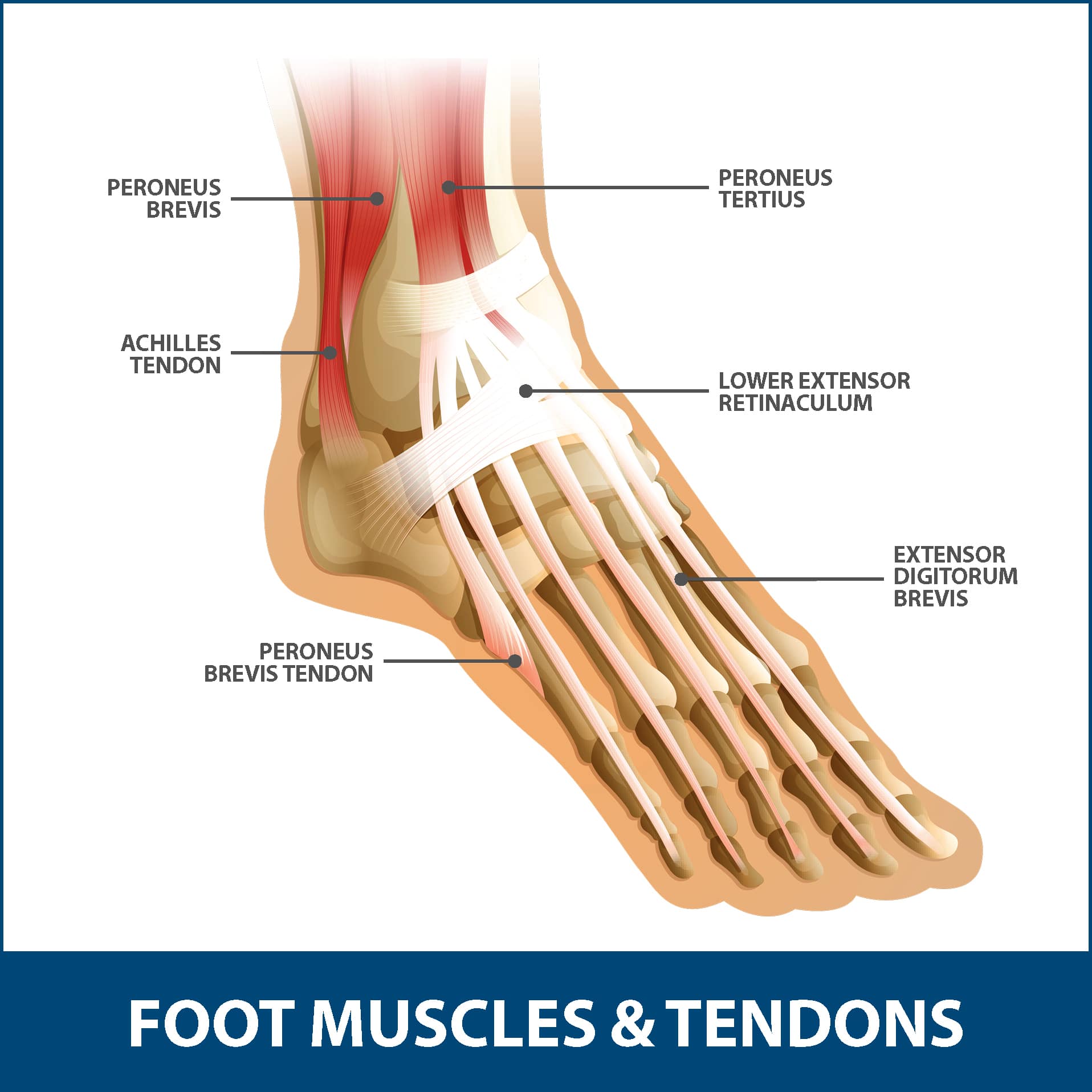
Anatomy Of Achilles Tendon
Its origin lies close to the middle of the calf and fuses with the gastrocnemius muscle proximally. Learn about the anatomy and vulernability to injury of the achilles tendon.
 Functional Anatomy Of The Achilles Tendon Springerlink
It is the tendinous extension of the three headed calf muscle consisting of soleus and the two headed gastrocnemius.

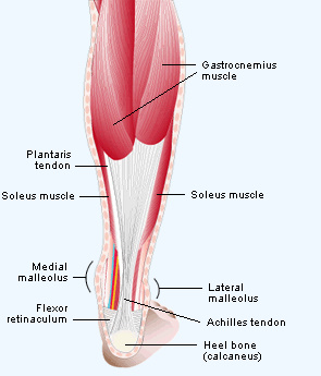
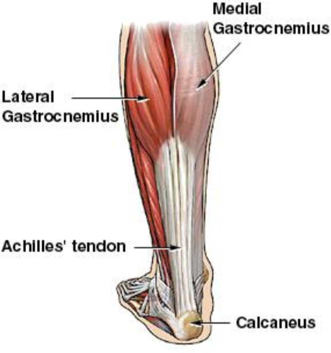
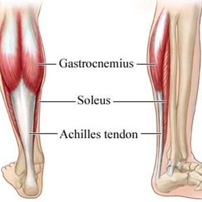
Anatomy of achilles tendon. Three relatively large and extremely strong muscles in the calf the gastrocnemius soleus and plantaris all attach to the back of the heel bone calcaneus via the achilles and the forces they generate during running and jumping are immense among the biggest in the body. The tendon is formed from the gastrocnemius and soleus muscles. The achilles tendon at is the thickest and strongest tendon in the human body.
It is named after the ancient greek mythological figure achilles. The plantaris tendon also fuses with the medial side of the achilles tendon proximal to its attachment site. The gastrocnemius is a fusiform muscle formed by two heads medial and lateral each separately crossing the knee joint.
Achilles tendon strong tendon at the back of the heel that connects the calf muscles to the heel. It is formed when the soleus muscle. The calcaneal tendon also known as the tendon of achilles is a posterior leg tendon a fibrous connective tissue that joins muscles in the back of the leg.
The tendon provides a distal attachment site for the gastrocnemius lateral and medial heads as well as the soleus muscles. The achilles tendon is also called the calcaneal tendon. Anatomy and importance of the achilles tendon the achilles tendon tendo calcaneus or tendo achillis is the thickest and strongest tendon in the human body.
The achilles tendon is a tough band of fibrous tissue that connects the calf muscles to the heel bone calcaneus. It inserts onto the posterior surface of the calcaneus heel bone. Essential in the flexion of the subtalar joint also known as the talocalcaneal joint in the ankle which exists between the calcaneus heel bone and the talus bone.
Anatomy of the achilles tendon the achilles tendon also known as the calcaneal tendon is a white fibrous cord located at the back of the ankle. The achilles tendon is one of the most robust tendons in the body and for good reason.
 Open Achilles Tendon Repair Foot And Ankle Operative

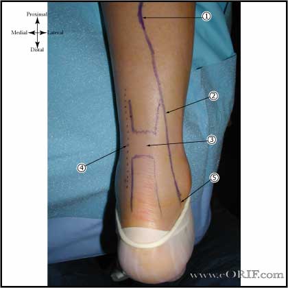 Achilles Tendon Repair Case Eorif
 Achilles Tendon Physiopedia
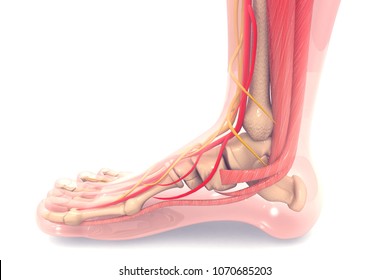
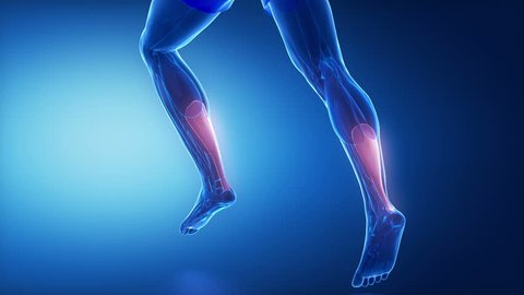 Achilles Tendon Leg Muscles Anatomy Animation
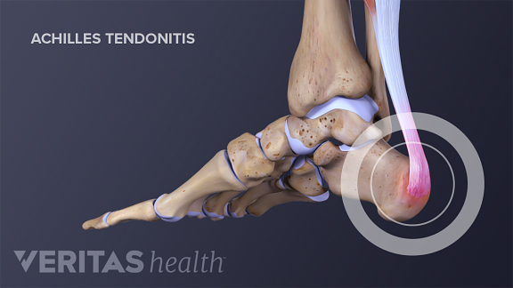
Achilles Tendinitis Middlesex Health
 Uncommon Injuries Sural Nerve Neuropathy
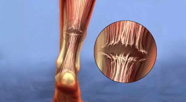
 Running Injuries Simplified Achilles Tendinopathy
 Achilles Tendon Tear Symptoms And Treatment Orthoinfo Aaos

 Ilustraciones Imagenes Y Vectores De Stock Sobre Achilles
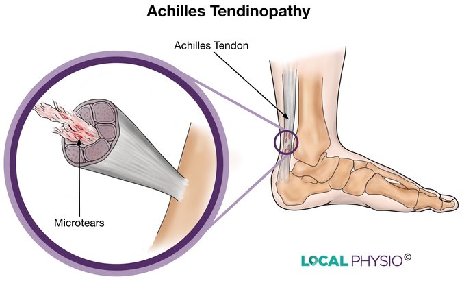 Achilles Tendon Local Physio
 Achilles Tendonitis And Tendon Injuries
 Achilles Tendon Physiology Achilles Tendon
 Are You Suffering From Achilles Tendon Pain Specialized

Achilles Tendon Anatomy And Importance
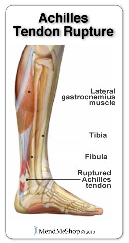
 Achilles Tendon Rupture Wikipedia
 Achilles Tendon Rupture Info Florida Orthopaedic Institute
 The Arterial Anatomy Of The Achilles Tendon Anatomical
 Achilles And Heel Pain Diagnosis Guide Physioadvisor
 Why Are Achilles Tendon Injuries So Common In Athletes
 Matles Test Physiopedia
 Achilles Tendon Rupture Core Em
 Injury To The Achilles Tendon George Herald
 In Vivo Mechanical Properties Of The Human Achilles Tendon
Belum ada Komentar untuk "Anatomy Of Achilles Tendon"
Posting Komentar